. 2015 Nov 25;10(11):e0143284.
doi: 10.1371/journal.pone.0143284. eCollection 2015.

Expression Patterns and Potential Biological Roles of Dip2a

Affiliations

Expression Patterns and Potential Biological Roles of Dip2a

Luqing Zhang et al. PLoS One. .

Abstract

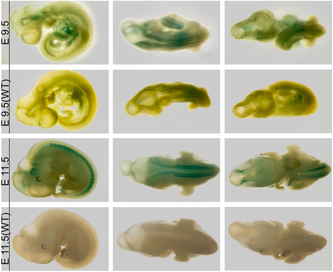
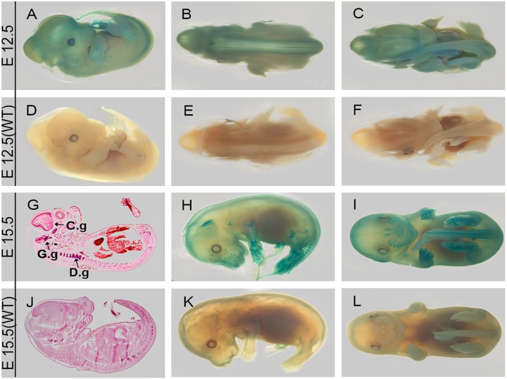
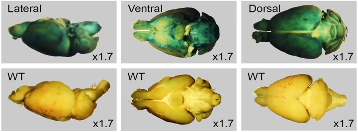
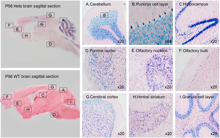
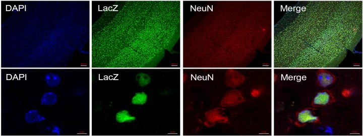
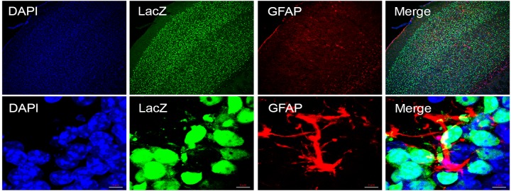
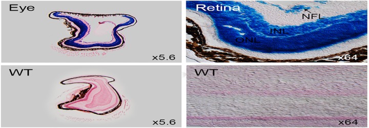
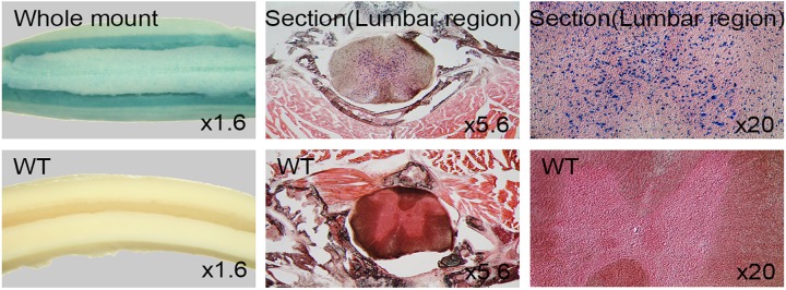
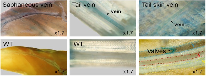
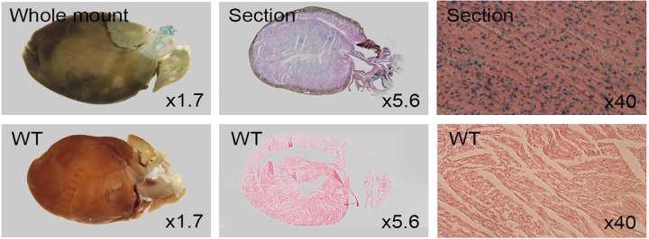
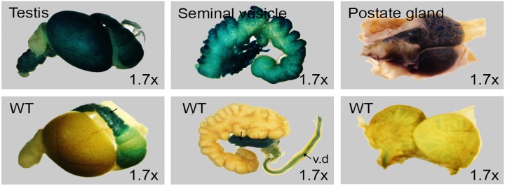
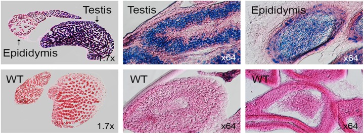
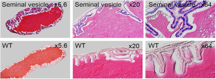
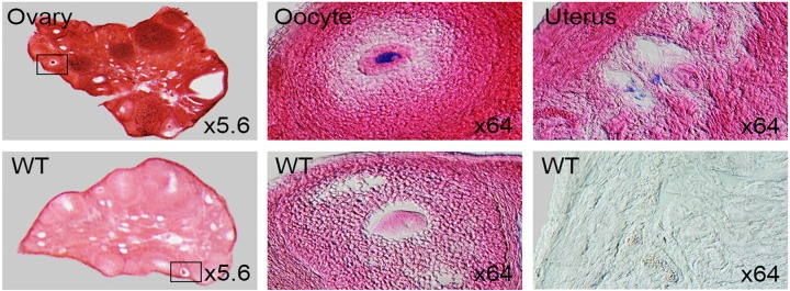
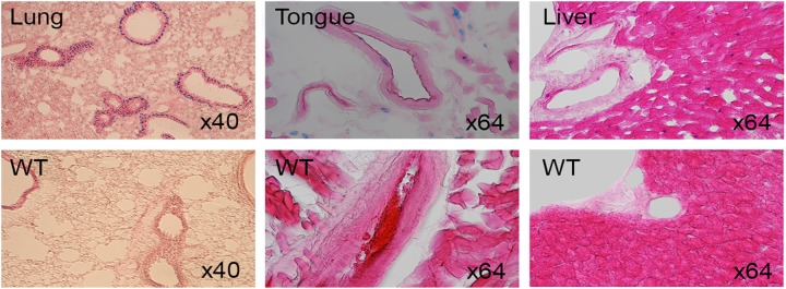
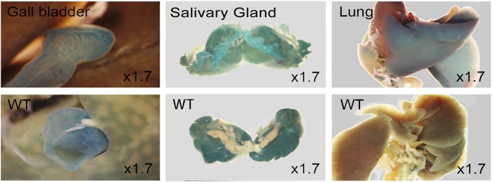
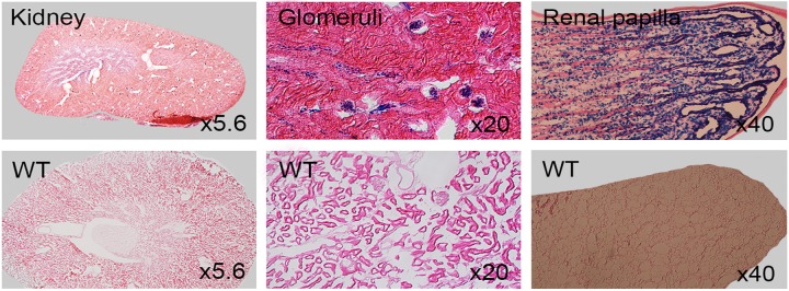
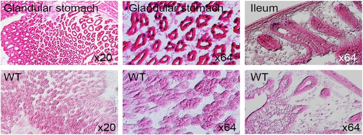
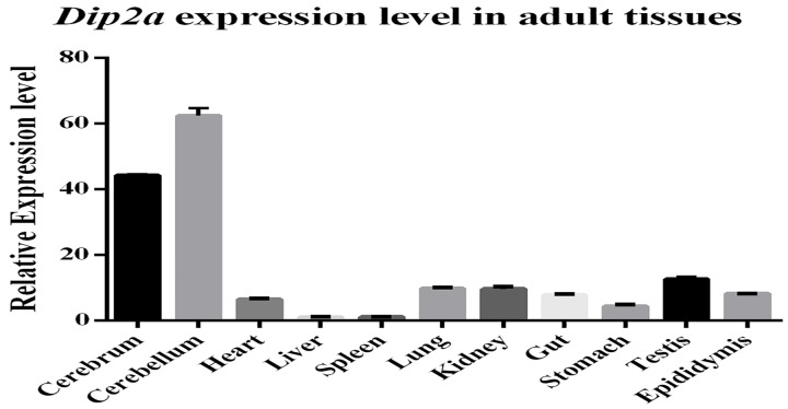
Disconnected (disco)-interacting protein 2 homolog A is a member of the DIP2 protein family encoded by Dip2a gene. Dip2a expression pattern has never been systematically studied. Functions of Dip2a in embryonic development and adult are not known. To investigate Dip2a gene expression and function in embryo and adult, a Dip2a-LacZ mouse model was generated by insertion of β-Gal cDNA after Dip2a promoter using CRISPR/Cas9 technology. Dip2a-LacZ mouse was designed to be a lacZ reporter mouse as well as a Dip2a knockout mouse. Heterozygous mice were used to study endogenous Dip2a expression and homozygotes to study DIP2A-associated structure and function. LacZ staining indicated that Dip2a is broadly expressed in neuronal, reproductive and vascular tissues, as well as in heart, kidney, liver and lung. Results demonstrate that Dip2a is expressed in ectoderm-derived tissues in developing embryos. Adult tissues showed rich staining in neurons, mesenchymal, endothelial, smooth muscle cells and cardiomyocytes by cell types. The expression pattern highly overlaps with FSTL1 and supports previous report that DIP2A to be potential receptor of FSTL1 and its protective roles of cardiomyocytes. Broad and intense embryonic and adult expression of Dip2a has implied their multiple structural and physiological roles.


Conflict of interest statement

Competing Interests: The authors have declared that no competing interests exist.

Figures

Fig 1
Fig 1. E9.5 and E11.5 LacZ staining.
Control wild type embryos are labeled (WT).
Fig 2
Fig 2. E12.5 and E15.5 LacZ staining.
Coronal section of E15.5 (G) showed signals in cranial ganglion (C.g), gasserian ganglion (G.g) and dorsal root ganglion (D.g).
Fig 3
Fig 3. Whole mount LacZ staining of 8 week old adult brain.
Control wild type labeled WT.
Fig 4
Fig 4. Sagittal sections of LacZ stained 8 week old adult brain.
Fig 5
Fig 5. Immunofluorescent staining of 8 week old adult brain section.
Cortex region of brain stained using anti-NeuN (Red), a marker specific to neuron, anti-LacZ (Green) and nucleus stained with DAPI (Blue). Top and bottom rows are low and high magnification respectively.
Fig 6
Fig 6. Immunofluorescent staining of adult brain section.
Glial cells were stained with anti-GFAP (red), anti-LacZ stained green and nucleus stained with DAPI (Blue). Top and bottom rows are low and high magnification respectively.
Fig 7
Fig 7. LacZ staining of 8 week old adult retina.
Sagittal sections of 8 week old adult eye are stained for LacZ expression. LacZ positive retina (top row) showed strong signals in optic nerve fiber layer (NFL), inner nuclear layer (INL) and outer nuclear layer (ONL).
Fig 8
Fig 8. LacZ staining of adult spinal cord.
Whole mount staining of 8 week old adult spinal cords (left column), coronal section of lumbar region (middle column) and high magnification of central canal indicate positive LacZ staining.
Fig 9
Fig 9. Vein staining of 8 week old adult Dip2a-LacZ mice.
Saphenous veins, tail veins, tail skin veins and valves were stained positive while control (WT) was negative.
Fig 10
Fig 10. LacZ staining of 8 week old adult heart.
Uniform staining in cardiomyocytes while control showed negative. Whole mount (left), section (middle) and high magnification (right).
Fig 11
Fig 11. Whole mount LacZ staining of 8 week old male reproductive organs.
Testis (left), seminal vesicle (middle) and prostate gland (right) stained in parallel with wild type littermates controls (bottom row). Non-specific staining seen in epididymis tail region (arrow) and vas deferens (v.d).
Fig 12
Fig 12. Coronal sections of LacZ-stained 8 week old male testis and epididymis.
WT represents controls.
Fig 13
Fig 13. Coronal sections of 8 week old male LacZ-stained seminal vesicle.
WT represents controls.
Fig 14
Fig 14. Coronal sections of 8 week old LacZ-stained female reproductive organs.
WT represents controls where wild type uterus was not counter stained with Eosin.
Fig 15
Fig 15. Section of LacZ-stained 8 week old adult lung, tongue and liver.
WT represents wild type control.
Fig 16
Fig 16. Whole mount staining of 8 week old mice gall bladder, salivary gland and lung.
WT represents controls. Endogenous β-Galactosidase activity was observed in the gall bladder and salivary gland.
Fig 17
Fig 17. Coronal sections of 8 week old adult LacZ-stained kidney.
WT represents controls.
Fig 18
Fig 18. Coronal sections of LacZ-stained 8 week old adult stomach and gut.
WT represents controls.
Fig 19
Fig 19. Real-time PCR from total RNA of 8 week old tissues.
Relative mRNA levels were calculated using ΔΔCt method. Data shown are representative of at least 3 independent repeats and expressed as the mean ± s.d.

References

    1. Tanaka M, Murakami K, Ozaki S, Imura Y, Tong XP, Watanabe T, et al. DIP2 disco-interacting protein 2 homolog A (Drosophila) is a candidate receptor for follistatin-related protein/follistatin-like 1—analysis of their binding with TGF-beta superfamily proteins. FEBS J. 2010;277(20):4278–89. 10.1111/j.1742-4658.2010.07816.x - DOI - PubMed
    1. Mukhopadhyay M, Pelka P, DeSousa D, Kablar B, Schindler A, Rudnicki MA, et al. Cloning, genomic organization and expression pattern of a novel Drosophila gene, the disco-interacting protein 2 (dip2), and its murine homolog. Gene. 2002;293(1–2):59–65. - PubMed
    1. Cooper MA, Zhou R. beta-Galactosidase staining of lacZ fusion proteins in whole tissue preparations. Methods Mol Biol. 2013;1018:189–97. 10.1007/978-1-62703-444-9_18 - DOI - PubMed
    1. Zhang L, Jia R, Palange NJ, Satheka AC, Togo J, An Y, et al. Large genomic fragment deletions and insertions in mouse using CRISPR/Cas9. PLoS One. 2015;10(3):e0120396 10.1371/journal.pone.0120396 - DOI - PMC - PubMed
    1. Malumbres M, Mangues R, Ferrer N, Lu S, Pellicer A. Isolation of high molecular weight DNA for reliable genotyping of transgenic mice. Biotechniques. 1997;22(6):1114–9. - PubMed

Publication types