. 2016 Mar;79(3):366-78.
doi: 10.1002/ana.24571. Epub 2016 Jan 28.

Mitochondrial DNA Depletion in Respiratory Chain-Deficient Parkinson Disease Neurons

Affiliations

Mitochondrial DNA Depletion in Respiratory Chain-Deficient Parkinson Disease Neurons

Anne Grünewald et al. Ann Neurol. 2016 Mar.

Abstract

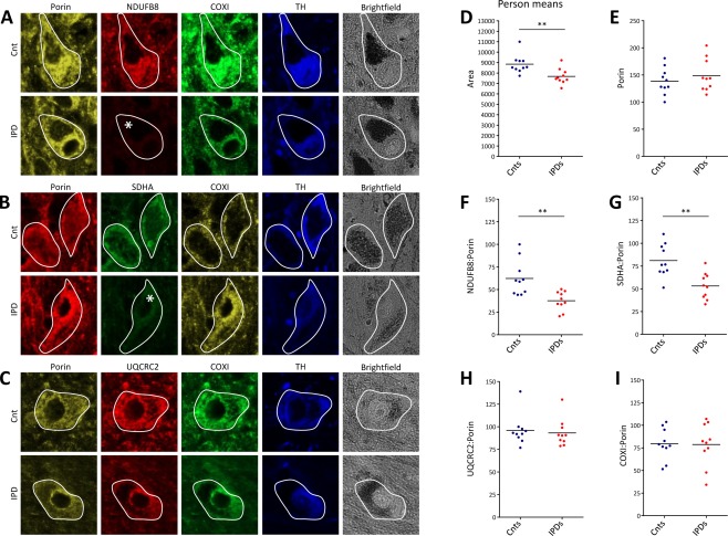
Objective: To determine the extent of respiratory chain abnormalities and investigate the contribution of mtDNA to the loss of respiratory chain complexes (CI-IV) in the substantia nigra (SN) of idiopathic Parkinson disease (IPD) patients at the single-neuron level.

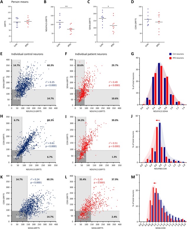
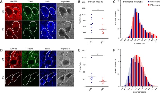
Methods: Multiple-label immunofluorescence was applied to postmortem sections of 10 IPD patients and 10 controls to quantify the abundance of CI-IV subunits (NDUFB8 or NDUFA13, SDHA, UQCRC2, and COXI) and mitochondrial transcription factors (TFAM and TFB2M) relative to mitochondrial mass (porin and GRP75) in dopaminergic neurons. To assess the involvement of mtDNA in respiratory chain deficiency in IPD, SN neurons, isolated with laser-capture microdissection, were assayed for mtDNA deletions, copy number, and presence of transcription/replication-associated 7S DNA employing a triplex real-time polymerase chain reaction (PCR) assay.

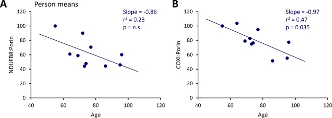
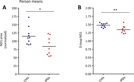
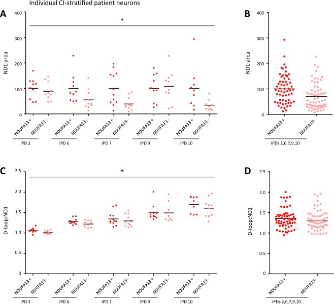
Results: Whereas mitochondrial mass was unchanged in single SN neurons from IPD patients, we observed a significant reduction in the abundances of CI and II subunits. At the single-cell level, CI and II deficiencies were correlated in patients. The CI deficiency concomitantly occurred with low abundances of the mtDNA transcription factors TFAM and TFB2M, which also initiate transcription-primed mtDNA replication. Consistent with this, real-time PCR analysis revealed fewer transcription/replication-associated mtDNA molecules and an overall reduction in mtDNA copy number in patients. This effect was more pronounced in single IPD neurons with severe CI deficiency.

Interpretation: Respiratory chain dysfunction in IPD neurons not only involves CI, but also extends to CII. These deficiencies are possibly a consequence of the interplay between nDNA and mtDNA-encoded factors mechanistically connected via TFAM.


Figures

Figure 1
Figure 1
Abundances of subunits of respiratory chain complexes CI–IV and mitochondrial mass in dopaminergic substantia nigra (SN) neurons from idiopathic Parkinson disease (IPD) patients and controls (Cnt). (A–C) Epifluorescence microscopy after quadruple‐label immunofluorescence indicated reduced levels of the CI subunit NDUFB8 and the CII subunit SDHA in IPD patient neurons, whereas the CIII subunit UQCRC2, the CIV subunit COXI, and the mitochondrial mass marker porin were unchanged. The investigated neurons were tyrosine hydroxylase (TH) ‐positive and contained neuromelanin deposits indicative of their dopaminergic character. Deficiencies are marked by an asterisk. (D) Despite their significantly smaller size, (E) densitometric analysis of area‐normalized porin levels indicated unchanged mitochondrial mass in patient neurons. In agreement with the results from the visual inspection, the average (F) NDUFB8:porin and (G) SDHA:porin ratios in IPD patient neurons were significantly reduced. By contrast, (H) UQCRC2:porin and (I) COXI:porin ratios were comparable in control and patient SN neurons. Lines indicate mean group ratios. **p < 0.01. COXI = cytochrome c oxidase subunit I; NDUFB8 = NADH dehydrogenase 1 beta subcomplex 8; SDHA = succinate dehydrogenase complex subunit A; UQCRC2 = ubiquinol–cytochrome c reductase core protein II.
Figure 2
Figure 2
Dependencies between respiratory chain complexes CI, CII, and CIV in single control (Cnt) and idiopathic Parkinson disease (IPD) patient substantia nigra (SN) neurons. (A–D) Densitometric analysis of the mitochondrial mass marker GRP75, the CI subunit NDUFA13, the CII subunit SDHA, and the CIV subunit COXI in patient and control SN neurons. Calculation of mean neuronal protein abundances per person confirmed reduced levels of mitochondria‐normalized CI and CII in IPD patients. (E) Spearman correlation analysis of NDUFA13:GRP75 and SDHA:GRP75 ratios indicated a weak link between the protein levels of both complexes in individual control neurons. The lower 25th percentile of the control ranges for NDUFA13:GRP75 and SDHA:GRP75 were used to define thresholds of deficiency (broken lines and shaded areas). (F) In patient neurons, the association between NDUFA13:GRP75 and SDHA:GRP75 was increased. Applying the deficiency thresholds, 49.1% of cells lacked both NDUFA13 and SDHA. Equal proportions of cells (10.6%) showed either isolated NDUFA13 or SDHA deficiency. (G) This caused overlapping frequency distributions of NDUFA13:SDHA in patient and control cells. (H, I) Spearman correlation analysis of NDUFA13:GRP75 and COXI:GRP75 revealed coregulation of CI and CIV in individual control and patient neurons. About a quarter of patient cells (25.3%) were deficient for NDUFA13 and COXI. In 34.3% of all patient neurons, isolated NDUFA13 deficiency occurred, whereas isolated COXI deficiency was only present in 1.3% of cells. (J) An overall decrease in NDUFA13 resulted in a shift of the NDUFA13:COXI ratio in IPD patient neurons relative to the control population. (K) Spearman correlation analysis of the SDHA:GRP75 and COXI:GRP75 ratios indicated a weak dependency between CII and CIV in control cells. (L) In patient neurons, this coregulation increased as a result of the combined loss of CII and CIV in 24.3% of cells. A large proportion of patient SN neurons (35.4%) also showed isolated SDHA deficiency, (M) causing the frequency distribution of SDHA:COXI to shift toward lower SDHA levels. *p < 0.05, **p < 0.01. COXI = cytochrome c oxidase subunit I; GRP75 = heat shock 70kDa protein 9 (mortalin); NDUFA13 = NADH dehydrogenase 1 alpha subcomplex 13; r 2 = Spearman correlation coefficient; SDHA = succinate dehydrogenase complex subunit A. [Color figure can be viewed in the online issue, which is available at www.annalsofneurology.org.]
Figure 3
Figure 3
Correlation of respiratory chain complex abundances in substantia nigra neurons of controls with age. Plotting of the average neuronal NDUFB8:porin (A) and COXI:porin (B) against age at death revealed age‐associated declines. COXI = cytochrome c oxidase subunit I; NDUFB8 = NADH dehydrogenase 1 beta subcomplex 8; n.s. = not significant; r 2 = Spearman correlation coefficient. [Color figure can be viewed in the online issue, which is available at www.annalsofneurology.org.]
Figure 4
Figure 4
Abundance of mtDNA‐associated TFAM and TFB2M in patient and control (Cnt) substantia nigra neurons. (A) Epifluorescence microscopy after triple‐label immunofluorescence indicated joint loss of NDUFB8 and TFAM in idiopathic Parkinson disease (IPD) patients, whereas porin staining showed normal mitochondrial mass. (B) Densitometric analysis of investigated proteins confirmed significantly reduced TFAM:porin ratios in IPD patients. Lines represent mean group ratios. (C) In patients and controls, NDUFB8 and TFAM strongly correlated at the single‐neuron level, resulting in similar behaviors of the frequency histograms for NDUFB8:TFAM. (D) Triple‐label immunohistochemistry showed reduced abundances of NDUFB8 and TFB2M but normal porin levels in IPD neurons. (E) Densitometric analysis of average neuronal TFB2M and porin levels in patients and controls revealed a significant reduction of TFB2M:porin in the former. (F) The frequency distribution of the NDUFB8:TFB2M ratio in single neurons showed a slight shift toward lower TFB2M in patient cells. *p < 0.05. NDUFB8 = NADH dehydrogenase 1 beta subcomplex 8; TFAM = mitochondrial transcription factor A; TFB2M = mitochondrial transcription factor B2.
Figure 5
Figure 5
mtDNA analysis in pooled substantia nigra neurons isolated from control (Cnt) and idiopathic Parkinson disease (IPD) patient midbrain sections. Simultaneous real‐time polymerase chain reaction quantification of mtDNA fragments in ND1 and the D‐loop in 30 neurons per person and experiment from 10 controls and 9 IPD patients are shown. This analysis indicated significantly reduced (A) mtDNA copy numbers (ND1:area) and (B) D‐loop:ND1 ratios in IPD patients compared to controls. *p < 0.05, **p < 0.01. ND1 = NADH dehydrogenase 1. [Color figure can be viewed in the online issue, which is available at www.annalsofneurology.org.]
Figure 6
Figure 6
mtDNA real‐time polymerase chain reaction analysis in individually isolated patient substantia nigra neurons with high or low CI abundance. (A) Determination of the ND1:area ratio in single NDUFA13‐positive and NDUFA13‐negative neurons from 5 idiopathic Parkinson disease (IPD) patients showed significantly reduced mtDNA copy numbers in the latter cells. (B) On average, NDUFA13‐negative patient neurons contained 29.4% fewer mtDNA copies than NDUFA13‐positive patient neurons. (C, D) Impaired mtDNA transcription/replication was indicated by a significant reduction in the D‐loop:ND1 ratio in NDUFA13‐negative neurons of all patients. *p < 0.05. ND1 = NADH dehydrogenase 1; NDUFA13 = NADH dehydrogenase 1 alpha subcomplex 13. [Color figure can be viewed in the online issue, which is available at www.annalsofneurology.org.]

References

    1. de Rijk MC, Tzourio C, Breteler MM, et al. Prevalence of parkinsonism and Parkinson's disease in Europe: the EUROPARKINSON Collaborative Study. European Community Concerted Action on the Epidemiology of Parkinson's Disease. J Neurol Neurosurg Psychiatry 1997;62:10–15. - PMC - PubMed
    1. Schapira AH. Mitochondrial pathology in Parkinson's disease. Mt Sinai J Med 2011;78:872–881. - PubMed
    1. Langston JW, Ballard P, Tetrud JW, Irwin I. Chronic parkinsonism in humans due to a product of meperidine‐analog synthesis. Science 1983;219:979–980. - PubMed
    1. Schapira AH, Cooper JM, Dexter D, et al. Mitochondrial complex I deficiency in Parkinson's disease. Lancet 1989;1:1269. - PubMed
    1. Kumar KR, Djarmati‐Westenberger A, Grunewald A. Genetics of Parkinson's disease. Semin Neurol 2011;31:433–440. - PubMed

Publication types

Substances