. 2015 Nov 25;10(11):e0143593.
doi: 10.1371/journal.pone.0143593. eCollection 2015.

M2 Polarization of Human Macrophages Favors Survival of the Intracellular Pathogen Chlamydia pneumoniae

Affiliations

M2 Polarization of Human Macrophages Favors Survival of the Intracellular Pathogen Chlamydia pneumoniae

Tanja Buchacher et al. PLoS One. .

Abstract

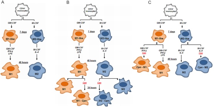
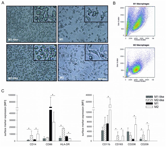
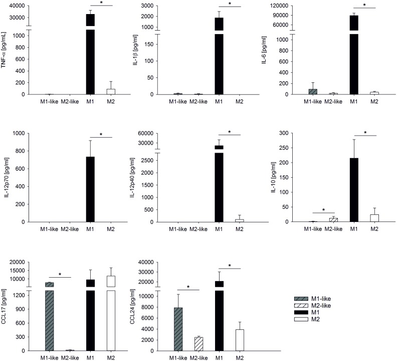
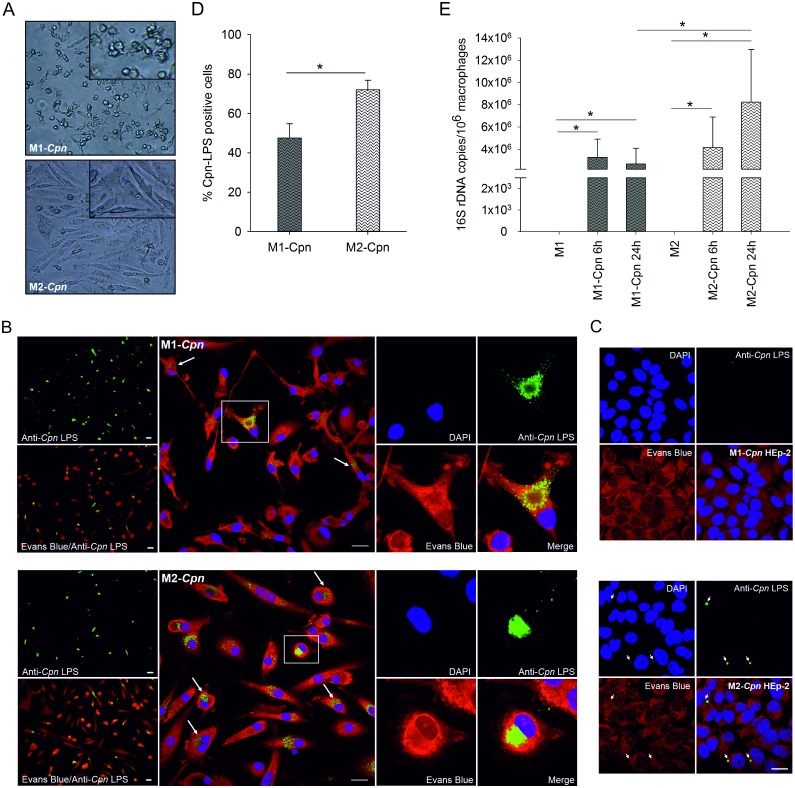
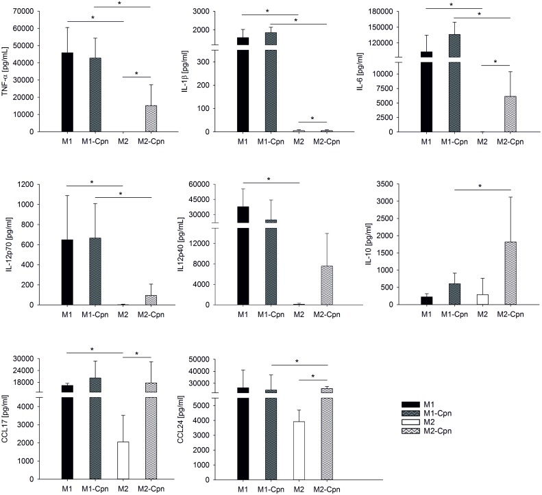
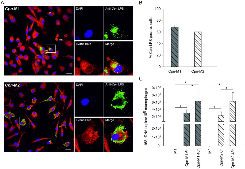
Intracellular pathogens have developed various strategies to escape immunity to enable their survival in host cells, and many bacterial pathogens preferentially reside inside macrophages, using diverse mechanisms to penetrate their defenses and to exploit their high degree of metabolic diversity and plasticity. Here, we characterized the interactions of the intracellular pathogen Chlamydia pneumoniae with polarized human macrophages. Primary human monocytes were pre-differentiated with granulocyte macrophage colony-stimulating factor or macrophage colony-stimulating factor for 7 days to yield M1-like and M2-like macrophages, which were further treated with interferon-γ and lipopolysaccharide or with interleukin-4 for 48 h to obtain fully polarized M1 and M2 macrophages. M1 and M2 cells exhibited distinct morphology with round or spindle-shaped appearance for M1 and M2, respectively, distinct surface marker profiles, as well as different cytokine and chemokine secretion. Macrophage polarization did not influence uptake of C. pneumoniae, since comparable copy numbers of chlamydial DNA were detected in M1 and M2 at 6 h post infection, but an increase in chlamydial DNA over time indicating proliferation was only observed in M2. Accordingly, 72±5% of M2 vs. 48±7% of M1 stained positive for chlamydial lipopolysaccharide, with large perinuclear inclusions in M2 and less clearly bordered inclusions for M1. Viable C. pneumoniae was present in lysates from M2, but not from M1 macrophages. The ability of M1 to restrict chlamydial replication was not observed in M1-like macrophages, since chlamydial load showed an equal increase over time for M1-like and M2-like macrophages. Our findings support the importance of macrophage polarization for the control of intracellular infection, and show that M2 are the preferred survival niche for C. pneumoniae. M1 did not allow for chlamydial proliferation, but failed to completely eliminate chlamydial infection, giving further evidence for the ability of C. pneumoniae to evade cellular defense and to persist in human macrophages.


Conflict of interest statement

Competing Interests: The authors have declared that no competing interests exist.

Figures

Fig 1
Fig 1. Macrophage Polarization.
(A) CD14 positive human blood monocytes were treated either with 25 ng/mL GM-CSF or 50 ng/mL M-CSF for 7 days to yield M1-like or M2-like macrophages. M1-like macrophages were activated with GM-CSF, LPS and 50 ng/mL IFN-γ for an additional 48 h to yield M1 macrophages, while M2-like macrophages were treated with M-CSF and IL-4 to yield M2. (B) To examine the impact of macrophage polarization on survival and proliferation of C. pneumoniae, M1 and M2 macrophages were infected and cultured with C. pneumoniae for 24 h. (C) M1-like macrophages were infected with C. pneumoniae and cultured for 48 h in the presence of GM-CSF and IFN-γ, while infected M2-like macrophages were cultured with M-CSF and IL-4. Uninfected macrophages served as control.
Fig 2
Fig 2. Morphology and surface marker expression of polarized macrophages.
Monocytes were cultured and polarized as shown in Scheme 1A to generate M1-like, M2-like, M1, and M2 macrophages. Morphology was assessed by light microscopy (panel A, scale bar 100 μm) and by flow cytometry according to forward/side scatter characteristics (panel B). The expression of surface markers was determined by flow cytometry (panel C). Data are expressed as mean ± SD for 3 independent experiments. MFI, mean fluorescence intensity.
Fig 3
Fig 3. Cytokine secretion of polarized macrophages.
Monocytes were cultured and polarized as shown in Scheme 1A to generate M1-like, M2-like, M1, and M2 macrophages. Cytokines were quantified in the culture supernatants of M1-like and M2-like macrophages after 7 days, and in the supernatants of M1 and M2 macrophages after an additional 48 h. Concentrations are expressed as mean ± SD for 3 independent experiments.
Fig 4
Fig 4. Visualization and quantification of C. pneumoniae in M1 and M2 macrophages.
Monocytes were cultured and polarized as shown in Scheme 1B and fully polarized M1 and M2 macrophages (4x10⁵/mL each) were infected with C. pneumoniae (4x10⁴ IFU) for 24 h. The morphology of infected M1 and M2 macrophages was determined by light microscopy (panel A, scale bar 100 μm). C. pneumoniae (green) was detected in M1 and M2 macrophages at 24 h post infection using immunofluorescence (panel B; scale bar = 20 μm). Cells were counterstained with Evans Blue (red), and DNA was visualized with DAPI (blue). Recovery of C. pneumoniae was evaluated by recultivating disrupted M1 and M2 macrophages 24 h post infection in HEp-2 cells (panel C; scale bar = 20 μm). The percentage of M1 and M2 macrophages that stained positive for C. pneumoniae at 24 h post infection was calculated by counting a minimum of 100 cells per slide (panel D). C. pneumoniae 16S rDNA was quantified by real-time PCR in infected M1 and M2 macrophages and in uninfected cells at 6 and 24 h post infection (panel E). Data are expressed as mean ± SD for 3 independent experiments.
Fig 5
Fig 5. Cytokine release of M1 and M2 macrophages infected with C. pneumoniae.
Monocytes were cultured and polarized as shown in Scheme 1B and fully polarized M1 and M2 macrophages (4x10⁵/mL each) were infected with C. pneumoniae (4x10⁴ IFU) for 24 h. Cytokine concentrations are expressed as mean ± SD for 3 independent experiments.
Fig 6
Fig 6. Detection of C. pneumoniae in M1-like and M2-like macrophages.
M1-like and M2-like macrophages (4x10⁵/mL each) were polarized as shown in Scheme 1C and were infected with C. pneumoniae (4x10⁴ IFU) for 24 h. C. pneumoniae (green) was detected in M1-like (panel A, top) and M2-like macrophages (panel A, bottom) at 48 h post infection using immunofluorescence (scale bar = 20 μm). Cells were counterstained with Evans Blue (red), and DNA was visualized with DAPI (blue). The percentage of infected M1-like and M2-like macrophages at 48 h post infection was calculated by counting a minimum of 100 cells per slide (panel B). C. pneumoniae 16S rDNA was quantified by real-time PCR in infected M1-like and M2-like macrophages and in uninfected cells at 6 and 48 hours post infection. Data are expressed as mean ± SD for 3 independent experiments.

References

    1. Price JV, Vance RE. The Macrophage Paradox. Immunity. 2014;41(5):685–93. 10.1016/j.immuni.2014.10.015 . - DOI - PubMed
    1. Thi EP, Lambertz U, Reiner NE. Sleeping with the enemy: how intracellular pathogens cope with a macrophage lifestyle. PLoS pathogens. 2012;8(3):e1002551 10.1371/journal.ppat.1002551 - DOI - PMC - PubMed
    1. Ray K, Marteyn B, Sansonetti PJ, Tang CM. Life on the inside: the intracellular lifestyle of cytosolic bacteria. Nature reviews Microbiology. 2009;7(5):333–40. 10.1038/nrmicro2112 . - DOI - PubMed
    1. Kern JM, Maass V, Maass M. Molecular pathogenesis of chronic Chlamydia pneumoniae infection: a brief overview. Clinical microbiology and infection: the official publication of the European Society of Clinical Microbiology and Infectious Diseases. 2009;15(1):36–41. 10.1111/j.1469-0691.2008.02631.x . - DOI - PubMed
    1. Saikku P. Seroepidemiology in Chlamydia pneumoniae—atherosclerosis association. European heart journal. 2002;23(4):263–4. 10.1053/euhj.2001.2913 . - DOI - PubMed

Publication types

LinkOut - more resources