

Clinical Validation of a Smartphone-Based Adapter for Optic Disc Imaging in Kenya
- PMID: 26606110
- PMCID: PMC5321504
- DOI: 10.1001/jamaophthalmol.2015.4625
Clinical Validation of a Smartphone-Based Adapter for Optic Disc Imaging in Kenya
Abstract
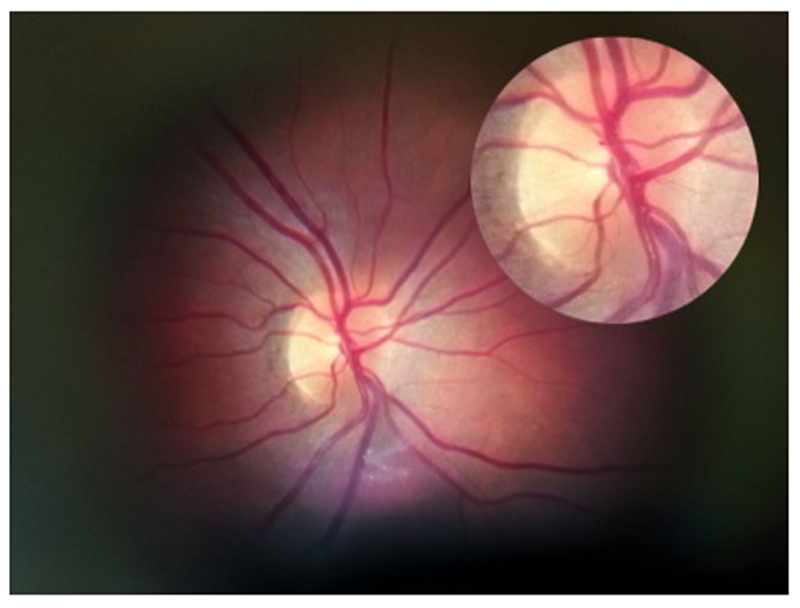
Importance: Visualization and interpretation of the optic nerve and retina are essential parts of most physical examinations.
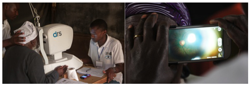
Objective: To design and validate a smartphone-based retinal adapter enabling image capture and remote grading of the retina.
Design, setting, and participants: This validation study compared the grading of optic nerves from smartphone images with those of a digital retinal camera. Both image sets were independently graded at Moorfields Eye Hospital Reading Centre. Nested within the 6-year follow-up (January 7, 2013, to March 12, 2014) of the Nakuru Eye Disease Cohort in Kenya, 1460 adults (2920 eyes) 55 years and older were recruited consecutively from the study. A subset of 100 optic disc images from both methods were further used to validate a grading app for the optic nerves. Data analysis was performed April 7 to April 12, 2015.
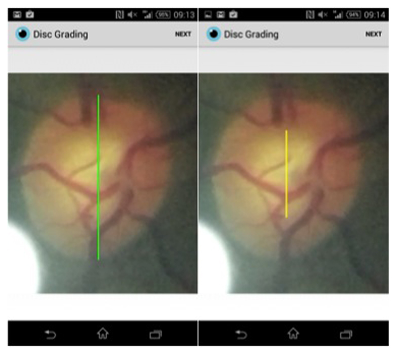
Main outcomes and measures: Vertical cup-disc ratio for each test was compared in terms of agreement (Bland-Altman and weighted κ) and test-retest variability.
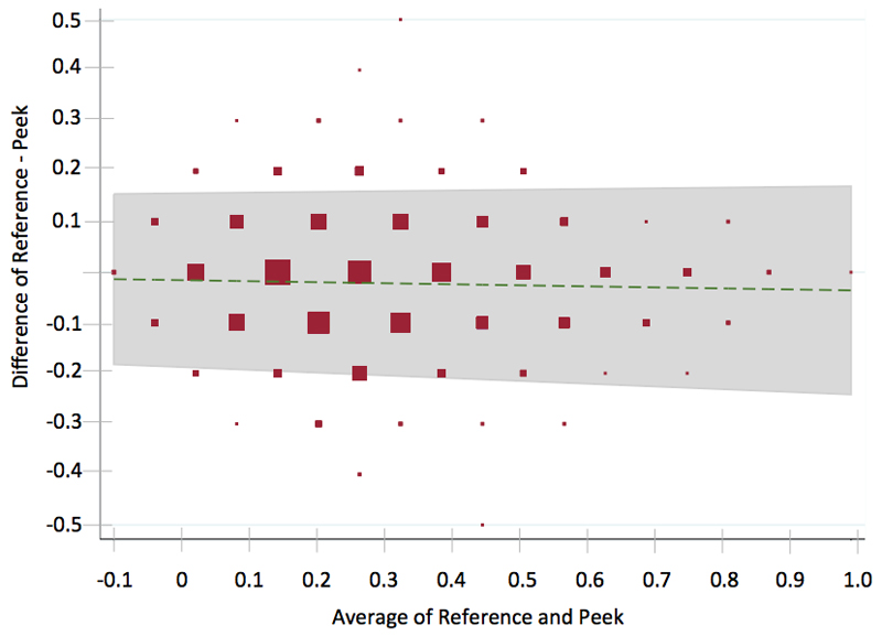
Results: A total of 2152 optic nerve images were available from both methods (also 371 from the reference camera but not the smartphone, 170 from the smartphone but not the reference camera, and 227 from neither the reference camera nor the smartphone). Bland-Altman analysis revealed a mean difference of 0.02 (95% CI, -0.21 to 0.17) and a weighted κ coefficient of 0.69 (excellent agreement). The grades of an experienced retinal photographer were compared with those of a lay photographer (no health care experience before the study), and no observable difference in image acquisition quality was found.
Conclusions and relevance: Nonclinical photographers using the low-cost smartphone adapter were able to acquire optic nerve images at a standard that enabled independent remote grading of the images comparable to those acquired using a desktop retinal camera operated by an ophthalmic assistant. The potential for task shifting and the detection of avoidable causes of blindness in the most at-risk communities makes this an attractive public health intervention.
Conflict of interest statement
Competing interests: All authors have completed the ICMJE uniform disclosure form at
The principal author affirms that this manuscript is an honest, accurate, and transparent account of the study being reported; that no important aspects of the study have been omitted; and that any discrepancies from the study as planned (and, if relevant, registered) have been explained.
The principal author had full access to all the data in the study and takes responsibility for the integrity of the data and the accuracy of the data analysis
Figures
Comment in
-
Applicability of Smartphone-Based Screening Programs.JAMA Ophthalmol. 2016 Feb;134(2):158-9. doi: 10.1001/jamaophthalmol.2015.4823. JAMA Ophthalmol. 2016. PMID: 26605498 No abstract available.
References
-
- Pascolini D, Mariotti SP. Global estimates of visual impairment: 2010. The British journal of ophthalmology. 2011 Dec 1; - PubMed
-
- Resnikoff S, Felch W, Gauthier TM, Spivey B. The number of ophthalmologists in practice and training worldwide: a growing gap despite more than 200 000 practitioners. The British journal of ophthalmology. 2012 Mar 26; - PubMed
-
- Scott KE, Kim DY, Wang L, et al. Telemedical diagnosis of retinopathy of prematurity intraphysician agreement between ophthalmoscopic examination and image-based interpretation. Ophthalmology. 2008 Jul;115(7):1222–1228 e1223. - PubMed
Publication types
MeSH terms
Substances
Grants and funding
LinkOut - more resources
Full Text Sources
Other Literature Sources
Medical
Miscellaneous
