

White Matter Changes Associated with Resting Sympathetic Tone in Frontotemporal Dementia vs. Alzheimer's Disease
- PMID: 26606247
- PMCID: PMC4659677
- DOI: 10.1371/journal.pone.0142445
White Matter Changes Associated with Resting Sympathetic Tone in Frontotemporal Dementia vs. Alzheimer's Disease
Abstract
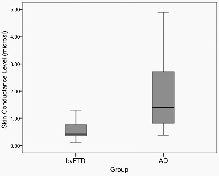
Background: Resting sympathetic tone, a measure of physiological arousal, is decreased in patients with apathy and inertia, such as those with behavioral variant frontotemporal dementia (bvFTD) and other frontally-predominant disorders.
Objective: To identify the neuroanatomical correlates of skin conductance levels (SCLs), an index of resting sympathetic tone and apathy, among patients with bvFTD, where SCLs is decreased, compared to those with Alzheimer's disease (AD), where it is not.
Methods: This study analyzed bvFTD (n = 14) patients and a comparison group with early-onset AD (n = 19). We compared their resting SCLs with gray matter and white matter regions of interest and white matter measures of fiber integrity on magnetic resonance imaging and diffusion tensor imaging.
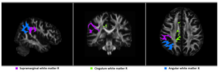
Results: As expected, bvFTD patients, compared to AD patients, had lower SCLs, which correlated with an apathy measure, and more gray matter loss and abnormalities of fiber integrity (fractional anisotropy and mean diffusivity) in frontal-anterior temporal regions. After controlling for group membership, the SCLs were significantly correlated with white matter volumes in the cingulum and inferior parietal region in the right hemisphere.
Conclusion: Among dementia patients, SCLs, and resting sympathetic tone, may correlate with quantity of white matter, rather than with gray matter or with white matter fiber integrity. Loss of white matter volumes, especially involving a right frontoparietal network, may reflect chronic loss of cortical axons that mediate frontal control of resting sympathetic tone, changes that could contribute to the apathy and inertia of bvFTD and related disorders.
Conflict of interest statement
Figures
Similar articles
-
Longitudinal white matter change in frontotemporal dementia subtypes and sporadic late onset Alzheimer's disease.Neuroimage Clin. 2017 Sep 14;16:595-603. doi: 10.1016/j.nicl.2017.09.007. eCollection 2017. Neuroimage Clin. 2017. PMID: 28975068 Free PMC article.
-
Exploring quantitative group-wise differentiation of Alzheimer's disease and behavioural variant frontotemporal dementia using tract-specific microstructural white matter and functional connectivity measures at multiple time points.Eur Radiol. 2019 Oct;29(10):5148-5159. doi: 10.1007/s00330-019-06061-7. Epub 2019 Mar 11. Eur Radiol. 2019. PMID: 30859283 Free PMC article.
-
Profiles of white matter tract pathology in frontotemporal dementia.Hum Brain Mapp. 2014 Aug;35(8):4163-79. doi: 10.1002/hbm.22468. Epub 2014 Feb 7. Hum Brain Mapp. 2014. PMID: 24510641 Free PMC article.
-
Joint assessment of white matter integrity, cortical and subcortical atrophy to distinguish AD from behavioral variant FTD: A two-center study.Neuroimage Clin. 2015 Sep 9;9:418-29. doi: 10.1016/j.nicl.2015.08.022. eCollection 2015. Neuroimage Clin. 2015. PMID: 26594624 Free PMC article.
-
Repetitive and stereotypic phenomena and dementia.Am J Alzheimers Dis Other Demen. 2013 May;28(3):223-7. doi: 10.1177/1533317513481094. Epub 2013 Mar 19. Am J Alzheimers Dis Other Demen. 2013. PMID: 23512997 Free PMC article. Review.
Cited by
-
The attribution of animacy and agency in frontotemporal dementia versus Alzheimer's disease.Cortex. 2017 Jul;92:81-94. doi: 10.1016/j.cortex.2017.03.019. Epub 2017 Apr 8. Cortex. 2017. PMID: 28458182 Free PMC article.
-
A novel method for objective quantification of apathy based on gaze and physiological reactivity to stimuli presented in a virtual reality environment.Alzheimers Dement (Amst). 2025 Jan 14;17(1):e70020. doi: 10.1002/dad2.70020. eCollection 2025 Jan-Mar. Alzheimers Dement (Amst). 2025. PMID: 40061180 Free PMC article.
-
"Lambda-wave" ST-elevation is associated with severe prognosis in stress (takotsubo) cardiomyopathy.Ann Noninvasive Electrocardiol. 2018 Nov;23(6):e12581. doi: 10.1111/anec.12581. Epub 2018 Jul 9. Ann Noninvasive Electrocardiol. 2018. PMID: 29984535 Free PMC article.
-
Brain tissue integrity mapping in adults with obstructive sleep apnea using T1-weighted and T2-weighted images.Ther Adv Neurol Disord. 2022 Nov 19;15:17562864221137505. doi: 10.1177/17562864221137505. eCollection 2022. Ther Adv Neurol Disord. 2022. PMID: 36419869 Free PMC article.
-
Autonomic dysfunction in neurodegenerative disease.Nat Rev Neurosci. 2025 May;26(5):276-292. doi: 10.1038/s41583-025-00911-8. Epub 2025 Mar 26. Nat Rev Neurosci. 2025. PMID: 40140684 Review.
References
-
- Srikanth S, Nagaraja AV, Ratnavalli E (2005) Neuropsychiatric symptoms in dementia-frequency, relationship to dementia severity and comparison in Alzheimer’s disease, vascular dementia and frontotemporal dementia. J Neurol Sci 236: 43–48. - PubMed
-
- Marin RS (1991) Apathy: a neuropsychiatric syndrome. J Neuropsychiatry Clin Neurosci 3: 243–254. - PubMed
-
- Levy R, Dubois B (2006) Apathy and the functional anatomy of the prefrontal cortex-basal ganglia circuits. Cereb Cortex 16: 916–928. - PubMed
Publication types
MeSH terms
Grants and funding
LinkOut - more resources
Full Text Sources
Other Literature Sources
Medical
Research Materials
