. 2016 Jan 5;88(1):890-7.
doi: 10.1021/acs.analchem.5b03536. Epub 2015 Dec 9.

A Dynamic Model of pH-Induced Protein G'e Higher Order Structure Changes derived from Mass Spectrometric Analyses

Affiliations

A Dynamic Model of pH-Induced Protein G'e Higher Order Structure Changes derived from Mass Spectrometric Analyses

Yelena Yefremova et al. Anal Chem. .

Abstract

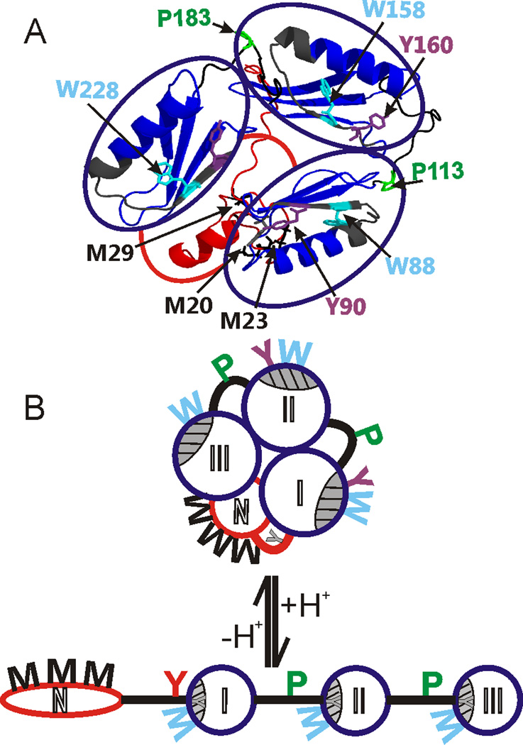
To obtain insight into pH change-driven molecular dynamics, we studied the higher order structure changes of protein G'e at the molecular and amino acid residue levels in solution by using nanoESI- and IM-mass spectrometry, CD spectroscopy, and protein chemical modification reactions (protein footprinting). We found a dramatic change of the overall tertiary structure of protein G'e when the pH was changed from neutral to acidic, whereas its secondary structure features remained nearly invariable. Limited proteolysis and surface-topology mapping of protein G'e by fast photochemical oxidation of proteins (FPOP) under neutral and acidic conditions reveal areas where higher order conformational changes occur on the amino-acid residue level. Under neutral solution conditions, lower oxidation occurs for residues of the first linker region, whereas greater oxidative modifications occur for amino-acid residues of the IgG-binding domains I and II. We propose a dynamic model of pH-induced structural changes in which protein G'e at neutral pH adopts an overall tight conformation with all four domains packed in a firm assembly, whereas at acidic pH, the three IgG-binding domains form an elongated alignment, and the N-terminal, His-tag-carrying domain unfolds. At the same time the individual IgG-binding domains themselves seem to adopt a more compacted fold. As the secondary structure features are nearly unchanged at either pH, interchange between both conformations is highly reversible, explaining the high reconditioning power of protein G'e-based affinity chromatography columns.


Figures

Figure 1
Figure 1
Ion mobility arrival times plotted as functions of charge states and nanoESI mass-spectra of protein G´e. A, B: Protein G´e was sprayed from 2% acetic acid:MeOH (95:5), pH 2.5. The drift time distribution for the 21+ ion (m/z 1239.09) is shown on the right. A series of narrow multiply charged ion signals with high charge numbers are recorded as doublets, indicating unfolded protein conformations. The satellite ion signal (on the right of the doublets) arises from gluconoylation. C, D: Protein G´e was sprayed from 50 mM ammonium acetate, pH 7. The drift time distribution for the 9+ ion (m/z 2909.82) is shown on the right. Broad ion signals with low charge numbers indicate globularly folded protein conformations.
Figure 2
Figure 2
Protein G´e amino acid sequence in single letter code. Oxidizable amino acid residues (M, Y, W, and P) are shown in bold. An N-terminal α-N-gluconoylation or α-N-6-phosphogluconoylation is indicated by “#”. Tn: numbers of tryptic peptides. Domains and higher order structure details are indicated with boxes and lines below the sequence. N: N-terminal region with His-tag; L: linker; I, II, III: IgG binding domains; S1, S2: spacer regions. Dashed boxes show regions that directly interact with the Fc parts of IgG. Fragments upon limited proteolysis with trypsin are dsignated “a”, “b” and “c” (cf. Supplemental Figure 4).
Figure 3
Figure 3
Chromatographic traces of nanoLC-separated tryptic peptides from protein G´e. A: After FPOP of protein G´e in 50 mM ammonium acetate, pH 7. B: After FPOP of protein G´e in 2% acetic acid, pH 2.5. Peak numbers with differential oxidation results between the two experiments are printed in bold.
Figure 4
Figure 4
Peak 11 of the nanoLC-ESI-MS run of protein G´e-derived tryptic peptides after FPOP in 50 mM ammonium acetate (cf. Figure 3A). A: The mass spectrum shows ions of m/z 1089.451 and 1097.447, which are assigned to peptides T9 and/or T15 with one and two oxidations, respectively. For ion signal assignments see supplemental table 5. B: MS/MS fragment ions from the precursor ion at m/z 1097.447. The corresponding amino acid sequence is shown in the insert and oxidation sites on W88 and/or W158 and Y90 and/or Y160 are depicted together with the fragment ion type assignments. B-type and Y"-type fragment ion signals are labeled; Y"n are labeled as Yn for simplicity.
Figure 5
Figure 5
A: Ribbon structure model of protein G´e. Domain assemblies at neutral pH. IgG-binding domains are circled in blue. Dark grey: IgG-binding surfaces. Red: N-terminal domain. Oxidized amino acid residues in the N-terminal domain, the three IgG binding domains, and linker and spacer peptides connecting the domains are shown in colored wireframes (black: M; purple: Y; cyan: W; green: P). B: Suggested models of protein G´e domain assemblies at neutral pH (top) and at acidic pH (bottom). Open circles represent the N-terminal domain (N; red), that is shown to be largely unstructured under acidic pH conditions, and the three IgG binding domains (I, II, III; blue), that maintain their secondary structure features under neutral and acidic conditions but become tighter at low pH. Thick lines represent linker (red) and spacer (black) peptides connecting the domains. Grey shaded and hatched areas depict IgG-binding surfaces. Amino acid residues (M, Y, W, P) are depicted to extrude from the structure cartoons when oxidized and buried in the interior when not oxidized (not accessible).

References

    1. Akerström B, Björck L. J. Biol. Chem. 1986;261:240–247. - PubMed
    1. Akerström B, Nielsen E, Björck L. J. Biol. Chem. 1987;262:13388–13391. - PubMed
    1. Alexander P, Fahnestock S, Lee T, Orban J, Bryan P. Biochemistry. 1992;31:3597–3603. - PubMed
    1. Guss B, Eliasson M, Olsson A, Uhlén M, Frej AK, Jörnvall H, Flock JI, Lindberg M. EMBO J. 1986;5:1567–1575. - PMC - PubMed
    1. Kronvall G. J. Immunol. 1973;111:1401–1406. - PubMed

Publication types