. 2015 Nov 25:15:170.
doi: 10.1186/s12886-015-0149-1.

Optical and material analysis of opacified hydrophilic intraocular lenses after explantation: a laboratory study

Affiliations

Optical and material analysis of opacified hydrophilic intraocular lenses after explantation: a laboratory study

Tamer Tandogan et al. BMC Ophthalmol. .

Abstract

Background: The opacification of hydrophilic intraocular lenses (IOLs) is a very rare complication in terms of absolute numbers. We report on the analyses of opacified Euromaxx ALI313Y and ALI313 IOLs (Argonoptics, Germany) using light and scanning electron microscopy, X-ray spectroscopy and optical bench analysis.

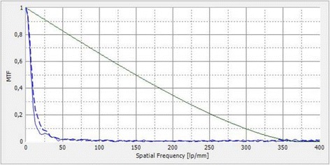
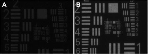
Methods: Opacified Euromaxx ALI313Y and ALI313 IOLs were explanted after patients presented with a decrease in visual acuity. The explants were sent to our laboratory and examined using light and scanning electron microscopy. The composition of the deposits was analysed using X-ray spectroscopy. The optical quality of the intraocular lens (IOL) was assessed using the OptiSpheric IOL PRO optical bench (Trioptics GmbH Wedel, Germany). Modulation transfer function (MTF) was measured at all spatial frequencies and United States Air Force (USAF) 1951 resolution target pictures were documented.

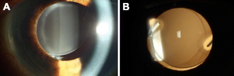
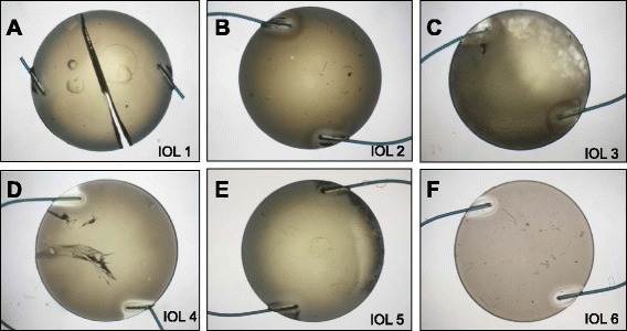
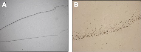
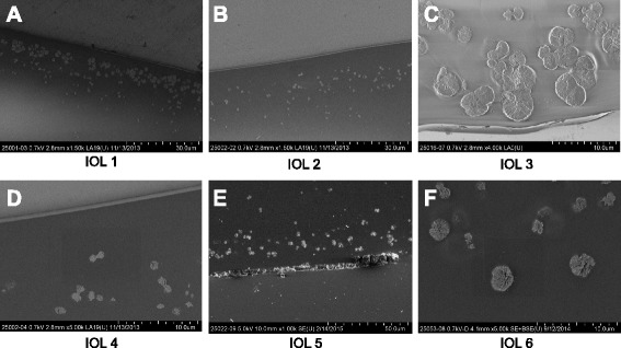
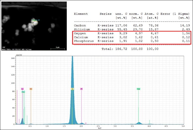
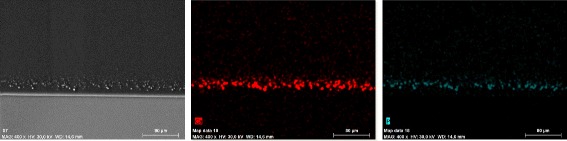
Results: Macroscopically, the entire optic was opacified in all IOLs. Light and scanning electron microscopy revealed numerous fine, granular, crystalline-like deposits, which were always distributed in a line parallel to the anterior and posterior surfaces of the IOLs. X-ray spectroscopy could prove the deposits consisted of Calcium and Phosphate. Measurements in the optical bench showed deterioration of MTF values at all spatial frequencies and the USAF target pictures demonstrated a significant reduction of brightness as well as resolution with the opacified IOLs.

Conclusions: The calcification of hydrophilic IOLs only occurs rarely. The exact chemical composition of the deposits can be assessed by means of X-ray spectroscopy. Optical quality analysis of the explanted Euromaxx ALI313Y and ALI313 IOLs showed significant reduction of MTF values, which was confirmed by USAF target pictures.


Figures

Fig. 1
Fig. 1
In vivo slit lamp image of a patient with an opacified ALI313 IOL. Focal a and retrograde b illumination
Fig. 2
Fig. 2
Gross examination of the opacified IOLs. a to e (IOL1 to 5): ALI313Y, f (IOL 6): ALI313
Fig. 3
Fig. 3
Exemplary light microscopy image of an opacified ALI313Y IOL. Staining with Alizarin Red
Fig. 4
Fig. 4
Light microscopy of the cross section of the IOLs analysed with von Kossa stain. Numerous fine, granular, crystalline-like deposits distributed in a line parallel to the anterior and posterior surface of IOLs. a Image showing both surfaces, b Image showing one surface in higher magnification
Fig. 5
Fig. 5
Scanning electron microscopy of the cross section of the IOLs. Numerous fine, granular, crystalline-like deposits distributed in a line parallel to the anterior (a, c, e) and posterior surface (b, d, f) of all IOLs. a to e (IOL 1 to 5) : ALI313Y, f (IOL 6) : ALI313
Fig. 6
Fig. 6
Example of X-ray spectroscopy in an opacified ALI313Y IOL. The deposits consist of Calcium phosphate. The Silicium-spike is an artefact caused by the Silicon-wafer, which is the substrate used for the analysis. Additionally, x-ray spectroscopy analysis on the surface of the IOLs (without Silicium-containing substrate) could exclude any Silicium within the precipitates
Fig. 7
Fig. 7
Example of element mapping in an opacified ALI313 IOL. Calcium is marked in red and phosphorus in green
Fig. 8
Fig. 8
An exemplary MTF analysis of an opacified ALI313Y IOL shows significant reduction of MTF values at all spatial frequencies. The green line shows the diffraction limit. The blue lines show the measured MTF values in sagittal and tangential planes
Fig. 9
Fig. 9
The USAF Target Analysis of the same IOL in Fig. 8. The image created by this IOL under standard light conditions shows a significant reduction of brightness and resolution a. Image created by the IOL with increased light b

Similar articles

Cited by

References

    1. Khoramnia R, Salgado JP, Auffarth GU, Schmidt S, Wegner A, Kobuch KA, Winkler von Mohrenfels C. [Opacification of a hydrophilic intraocular lens 4 years after cataract surgery. A biomaterial analysis] Ophthalmologe. 2012;109(5):483–486. doi: 10.1007/s00347-011-2487-6. - DOI - PubMed
    1. Fung SS, Sykakis E, Islam NM, Zambarakji HJ, Khoramnia R, Auffarth GU, Parmar DN. Intraocular Lens Opacification following Intracameral Injection of Recombinant Tissue Plasminogen Activator to Treat Inflammatory Membranes after Cataract Surgery. J Ophthalmol. 2015;2015:975075. doi: 10.1155/2015/975075. - DOI - PMC - PubMed
    1. Schmidinger G, Pemp B, Werner L. [Opacification of an intraocular lens: calcification of hydrophilic intraocular lenses after gas tamponade of the anterior chamber] Ophthalmologe. 2013;110(11):1066–1068. doi: 10.1007/s00347-013-2793-2. - DOI - PubMed
    1. Izak AM, Werner L, Pandey SK, Apple DJ. Calcification of modern foldable hydrogel intraocular lens designs. Eye. 2003;17(3):393–406. doi: 10.1038/sj.eye.6700341. - DOI - PubMed
    1. Werner L, Hunter B, Stevens S, Chew JJ, Mamalis N. Role of silicon contamination on calcification of hydrophilic acrylic intraocular lenses. Am J Ophthalmol. 2006;141(1):35–43. doi: 10.1016/j.ajo.2005.08.045. - DOI - PubMed

Publication types

LinkOut - more resources