. 2015 Nov 25:14:202.
doi: 10.1186/s12943-015-0473-3.

A three-miRNA signature as promising non-invasive diagnostic marker for gastric cancer

Affiliations

A three-miRNA signature as promising non-invasive diagnostic marker for gastric cancer

Vivian Yvonne Shin et al. Mol Cancer. .

Abstract

Background: Despite the declining incidence of gastric cancer, mortality rate remains high due to late presentation. We aimed to evaluate the sensitivity of miRNA as a diagnostic marker for gastric cancer in the circulation.

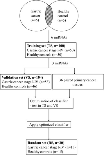
Methods: Plasma samples from 3 independent groups comprise 123 gastric cancer patients and 111 healthy controls for miRNA profiling from microarray screening.

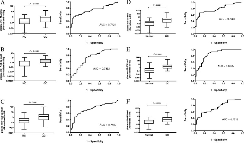
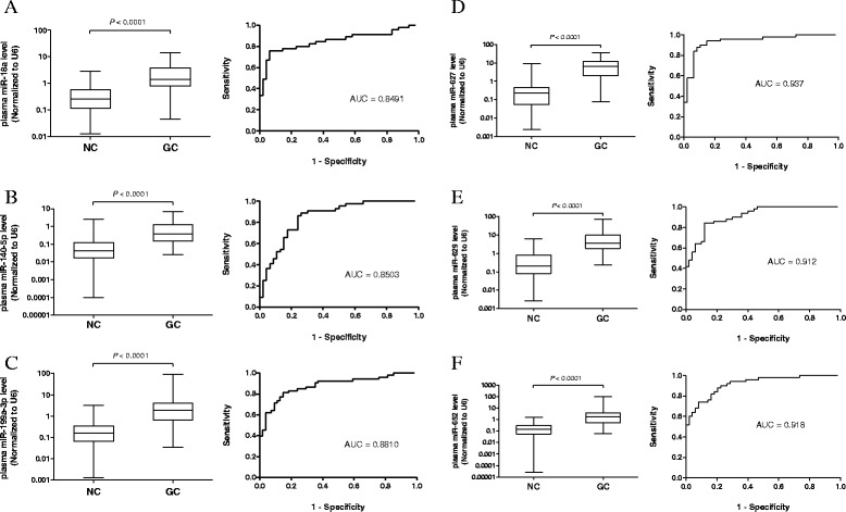
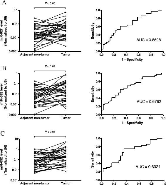
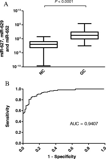
Results: Microarray data showed that 25 miRNAs were upregulated in gastric cancer patients and 6 highly expressed miRNAs (miR-18a, miR-140-5p, miR-199a-3p, miR-627, miR-629 and miR-652) were selected for validation. In an independent validation set, levels of miR-627, miR-629 and miR-652 were significantly higher in gastric cancer patients than healthy controls (P <0.0001). An algorithm with improved sensitivity and specificity as gastric cancer classifier was adopted and validated in another random set of 15 plasma samples. Results showed that combination of 3 miRNAs obtained the highest area under curve, with a cut-off at 0.373, with a sensitivity of 86.7% and a specificity of 85.5%.

Conclusion: This study revealed a three-miRNA signature as a promising classifier for gastric cancer, and greatly enhances the feasibility of circulating miRNAs as non-invasive diagnostic marker for this disease.


Figures

Fig. 1
Fig. 1
An overview of the workflow of the study design
Fig. 2
Fig. 2
Validation of plasma (a) miR-18a, (b) miR-140-5p, (c) 199a-3p, (d) miR-627, (e) miR-629 and (f) miR-652 levels in training set (TS). Expression levels were normalized to U6. Box plots of six miRNAs in plasma of gastric cancer patients (n = 50) and healthy controls (n = 50) The boxes mark the interval between the 25th and 75th percentiles, and the lines inside the box denote the medians. The whiskers represent the interval between the 10th and 90th percentiles. Statistically significant differences were analyzed using Mann–Whitney test. Receiver-operating characteristic (ROC) curve analysis of miRNA for discriminating gastric cancer patients from healthy controls
Fig. 3
Fig. 3
Selection and validation of (a) miR-18a, (b) miR-140-5p, (c) 199a-3p, (d) miR-627, (e) miR-629 and (f) miR-652 in an independent validation set (VS). Box plots and ROC curve analysis of miRNAs in the plasma of gastric cancer patients (n = 58) and healthy controls (n = 46). The boxes mark the interval between the 25th and 75th percentiles, and the lines inside the box denote the medians. The whiskers represent the interval between the 10th and 90th percentiles. Statistically significant differences were analyzed using Mann–Whitney test. Receiver-operating characteristic (ROC) curve analysis of miRNA for discriminating gastric cancer patients from healthy controls
Fig. 4
Fig. 4
Expression levels of (a) miR-627, (b) miR-629 and (c) miR-652 in paired gastric tumor tissues and adjacent non-tumor counterparts (n = 36). Expression levels were normalized to U6. Statistically significant differences were analyzed using Wilcoxon test. Receiver-operating characteristic (ROC) curve analysis of miRNA for discriminating gastric cancer patients from healthy controls
Fig. 5
Fig. 5
Validation of gastric cancer classifier in different validation sets. a Box plot and ROC curve analysis of combining 3 miRNAs in random set (RS) with gastric cancer patients (n = 15) and healthy controls (n = 15). b Box plot and ROC curve analysis of combining 3 miRNAs in TSVS with gastric cancer patients (n = 108) and healthy controls (n = 96). The boxes mark the interval between the 25th and 75th percentiles, and the lines inside the box denote the medians. The whiskers represent the interval between the 10th and 90th percentiles. Statistically significant differences were analyzed using Mann–Whitney test
Fig. 6
Fig. 6
Correlation between gastric cancer classifier and tumor stage in plasma of gastric cancer patients (n = 123)

References

    1. Bertuccio P, Chatenoud L, Levi F, Praud D, Ferlay J, Negri E, et al. Recent patterns in gastric cancer: a global overview. Int J Cancer. 2009;125:666–73. doi: 10.1002/ijc.24290. - DOI - PubMed
    1. Hundahl SA, Phillips JL, Menck HR. The National Cancer Data Base Report on poor survival of U.S. gastric carcinoma patients treated with gastrectomy: Fifth Edition American Joint Committee on Cancer staging, proximal disease, and the “different disease” hypothesis. Cancer. 2000;88:921–32. doi: 10.1002/(SICI)1097-0142(20000215)88:4<921::AID-CNCR24>3.0.CO;2-S. - DOI - PubMed
    1. Xia HH, Yang Y, Chu KM, Gu Q, Zhang YY, He H, et al. Serum macrophage migration-inhibitory factor as a diagnostic and prognostic biomarker for gastric cancer. Cancer. 2009;115:5441–9. doi: 10.1002/cncr.24609. - DOI - PubMed
    1. Chan AO, Lam SK, Chu KM, Lam CM, Kwok E, Leung SY, et al. Soluble E-cadherin is a valid prognostic marker in gastric carcinoma. Gut. 2001;48:808–11. doi: 10.1136/gut.48.6.808. - DOI - PMC - PubMed
    1. Chan AO, Chu KM, Lam SK, Cheung KL, Law S, Kwok KF, et al. Early prediction of tumor recurrence after curative resection of gastric carcinoma by measuring soluble E-cadherin. Cancer. 2005;104:740–6. doi: 10.1002/cncr.21260. - DOI - PubMed

Publication types