Review
. 2015 Nov 25:46:137.
doi: 10.1186/s13567-015-0266-0.

Amphibian chytridiomycosis: a review with focus on fungus-host interactions

Affiliations
Review

Amphibian chytridiomycosis: a review with focus on fungus-host interactions

Pascale Van Rooij et al. Vet Res. .

Abstract

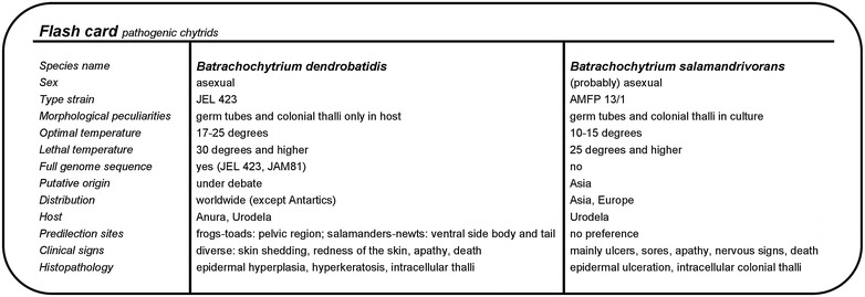
Amphibian declines and extinctions are emblematic for the current sixth mass extinction event. Infectious drivers of these declines include the recently emerged fungal pathogens Batrachochytrium dendrobatidis and Batrachochytrium salamandrivorans (Chytridiomycota). The skin disease caused by these fungi is named chytridiomycosis and affects the vital function of amphibian skin. Not all amphibians respond equally to infection and host responses might range from resistant, over tolerant to susceptible. The clinical outcome of infection is highly dependent on the amphibian host, the fungal virulence and environmental determinants. B. dendrobatidis infects the skin of a large range of anurans, urodeles and caecilians, whereas to date the host range of B. salamandrivorans seems limited to urodeles. So far, the epidemic of B. dendrobatidis is mainly limited to Australian, neotropical, South European and West American amphibians, while for B. salamandrivorans it is limited to European salamanders. Other striking differences between both fungi include gross pathology and thermal preferences. With this review we aim to provide the reader with a state-of-the art of host-pathogen interactions for both fungi, in which new data pertaining to the interaction of B. dendrobatidis and B. salamandrivorans with the host's skin are integrated. Furthermore, we pinpoint areas in which more detailed studies are necessary or which have not received the attention they merit.


Figures

Fig. 1
Fig. 1
Phylogeny and classification of the genus Batrachochytrium. Cladogram showing the taxonomic position of Batrachochytrium dendrobatidis and Batrachochytrium salamandrivorans within the fungal kingdom (a), the phylum Chytridiomycota (b) and order of the Rhizophydiales (c). The position of the Microsporidia remains uncertain. Branch lengths are not proportional to genetic distances. The topology is derived from Martel et al. [13], Longcore et al. [16] and Hibbett et al. [150]
Fig. 2
Fig. 2
Morphology of Batrachochytrium species in culture. a Culture of B. dendrobatidis on tryptone/gelatin-hydrolysate/lactose (TGhL)-broth, showing abundant mature zoosporangia (black arrow) containing zoospores and empty, discharged sporangia (white arrow); b In culture (TGhL-broth) B. salamandrivorans is characterized by predominant monocentric thalli (black arrow), few colonial thalli (white arrow) and zoospore cysts with germ tubes (asterisk); scale bars 100 µm
Fig. 3
Fig. 3
The lifecycle of Batrachochytrium species in culture. In culture Batrachochytrium dendrobatidis continues the life cycle stages A–E, while in Batrachochytrium salamandrivorans additional life cycle stages B1-B2 are observed: (A) flagellated motile zoospores; (B) encysted zoospore; (B1) germling with germtube; (B2) transfer of the cell contents into a newly formed thallus; (C) zoospore cyst with rhizoids; (D) immature sporangium; (E) mature monocentric zoosporangium with discharge tube (at the right), colonial thallus containing several sporangia, each with their own discharge tube (at the left). Modified from Berger et al. [23]
Fig. 4
Fig. 4
Clinical signs and pathology associated with infection due to Batrachochytrium dendrobatidis. a Naturally infected moribund common midwife toad (Alytes obstetricans) showing abnormal posture (abduction hind legs) and loose sloughed skin; b section through the ventral skin (drink patch) of the same infected toad; infection is characterized by diffuse epidermal hyperkeratosis and hyperplasia combined with the presence of numerous zoosporangia at various stages of maturation; HE; scale bar 50 µm; c detail of intracellular septate zoosporangia; HE; scale bar 10 µm
Fig. 5
Fig. 5
Clinical signs and pathology associated with infection due to Batrachochytrium salamandrivorans. a a naturally infected fire salamander (Salamandra salamandra) found during a B. salamandrivorans-outbreak (Robertville, Belgium) showing several ulcers (white arrows) and excessive skin shedding; b extensive ulceration (white arrows) at the ventral side of an infected fire salamander; c skin section through an ulcer evidences abundant intracellular colonial thalli in all epidermal skin layers; immunohistochemical stain with polyclonal antibodies to B. dendrobatidis; scale bar 10 µm; d magnification of the intracellular colonial thalli from micrograph c; immunohistochemical stain; scale bar 10 µm
Fig. 6
Fig. 6
Chemotaxis of Batrachochytrium dendrobatidis toward free integumental sugars. The sugars α-d-mannose (Man), α-d-galactose (Gal), α-l-fucose (Fuc), β-d-N-acetylglucosamine (GluNAc), α-d-N-acetylgalactosamine (GalNAc), N-acetylneuraminic acid (NeuNAc) or sialic acid were tested as attractans at a 0.1 M concentration, using a traditional capillary tube test. Water was used as vehicle and controle attractans. Genomic equivalents (GE) of B. dendrobatidis zoospores in the capillaries were quantified after a 90 min using quantitative real-time PCR. Mean ± standard error of three independent experiments are presented
Fig. 7
Fig. 7
Zoosporicidal activity of Xenopus laevis skin mucus at physiological concentrations. Killing activity is expressed as log(10) viable spores added to the skin secretions—log(10) viable spores recovered after 2 and 24 h incubation. Results are presented as mean genomic equivalents of B. dendrobatidis ±standard error (SEM). Sample size (n) for time point (T) = 3
Fig. 8
Fig. 8
Adhesion of Batrachochytrium dendrobatidis to Xenopus laevis skin. Adhesion to the epidermal surface is established both by tubular projections, possibly adhesins (black arrow) and rhizoids (white arrow). Some encysted zoospores have collapsed (asterisk) due to cell hollowing; scale bar 5 µm
Fig. 9
Fig. 9
Infection cycle of Batrachochytrium dendrobatidis in a susceptible host. The endobiotic lifecycle includes successively germ tube mediated invasion, establishment of intracellular thalli, spread to the deeper skin layers, upward migration by the differentiating epidermal cell to finally release zoospores at the skin surface
Fig. 10
Fig. 10
Epibiotic lifecycle of Batrachochytrium dendrobatidis. The epibiotic lifecycle observed in skin explants of Xenopus laevis includes germ tube mediated invasion, outgrowth of a rhizoidal network, uptake of host cell cytoplasm as nutrient for the growing and maturing chytrid thallus upon the skin surface
Fig. 11
Fig. 11
Flash card of the pathogenic chytrid fungi. Overview of the key features and most striking dissimilarities between the pathogenic chytrid fungi B. dendrobatidis and B. salamandrivorans (2015)

References

    1. IUCN (2015) IUCN Red List of Threatened Species. Version 2015-3. http://www.iucnredlist.org. Accessed 19 Sept 2015
    1. Gascon C, Collins JP, Moore RD, Church DR, McKay JE, Mendelson JR., III . Amphibian conservation action plan. Gland: IUCN/SSC Amphibian Specialist Group; 2007.
    1. Berger L, Speare R, Daszak P, Green DE, Cunningham AA, Goggin CL, Slocombe R, Ragan MA, Hyatt AD, McDonald KR, Hines HB, Lips KR, Marantelli G, Parkes H. Chytridiomycosis causes amphibian mortality associated with population declines in the rain forests of Australia and Central America. Proc Natl Acad Sci U S A. 1998;95:9031–9036. doi: 10.1073/pnas.95.15.9031. - DOI - PMC - PubMed
    1. Lips KR, Brem F, Brenes R, Reeve JD, Alford RA, Voyles J, Carey C, Livo L, Pessier AP, Collins JP. Emerging infectious disease and the loss of biodiversity in a Neotropical amphibian community. Proc Natl Acad Sci USA. 2006;103:3165–3170. doi: 10.1073/pnas.0506889103. - DOI - PMC - PubMed
    1. Campbell CR, Voyles J, Cook DI, Dinudom A. Frog skin epithelium: electrolyte transport and chytridiomycosis. Int J Biochem Cell Biol. 2012;44:431–434. doi: 10.1016/j.biocel.2011.12.002. - DOI - PMC - PubMed

Publication types

LinkOut - more resources