Review
. 2016 Apr 10;343(1):21-27.
doi: 10.1016/j.yexcr.2015.11.017. Epub 2015 Nov 24.

Mechanosensitive components of integrin adhesions: Role of vinculin

Affiliations
Review

Mechanosensitive components of integrin adhesions: Role of vinculin

Paul Atherton et al. Exp Cell Res. .

Abstract

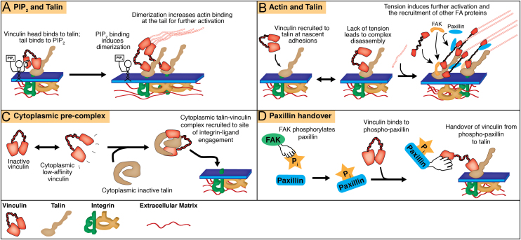
External forces play a key role in shaping development and normal physiology. Aberrant responses to forces, or changes in the nature of such forces, are implicated in a variety of diseases. Cells contain several types of adhesions, linking them to their external environment. It is through these adhesions that forces are both sensed (from the outside inwards) and applied (from inside to out). Furthermore, several adhesion-based proteins are sensitive to changes in intracellular forces, utilising them for activation and regulation. Here, we outline how vinculin, a key component of integrin-mediated adhesions linking the actin cytoskeleton to the extracellular matrix (ECM), is regulated by force and acts as force transducing protein. We discuss the role of vinculin in vivo and its place in health and disease; summarise the proposed mechanisms by which vinculin is recruited to and activated at integrin-ECM adhesions; and discuss recent findings that place vinculin as the major force sensing and transmitting component of cell-matrix adhesion complexes. Finally, we discuss the role of vinculin in regulating the cellular responses to both the physical properties of the external environment and to externally applied physical stimuli.

Keywords: Actin; Focal adhesion; Force; Mechanotransduction; Vinculin.


Figures

Fig. 1
Fig. 1
Models of vinculin recruitment and activation. Four potential mechanisms have been proposed to contribute to vinculin recruitment to integrin-ECM adhesion sites and activation. While these have been separated here, it is likely that a combination of these mechanisms is used in the cell. A. Vinculin binds to integrin-bound talin via the head domain. PIP2, which is enriched at these sites, binds to the vinculin tail leading to dimerization and increasing actin binding. B. Vinculin is recruited to talin bound to the cytoplasmic tail of integrin, inducing partial activation. Actin binding at the tail, providing actomyosin-based tension, is required for further activation of vinculin; without actin binding, the two proteins dissociate and the nascent adhesion does not mature. C. Vinculin undergoes rapid conformational changes in its tertiary structure, switching between an inactive and a low-affinity state. The low affinity state is able to bind to cytoplasmic talin (itself in either an inactive state, or also in a ‘low-affinity’ state (not shown)) to form a cytoplasmic ‘pre-complex’, which is then recruited to sites of integrin-ligand engagement. D. Paxillin is phosphorylated by FAK at nascent adhesions. Vinculin binds to phosphorylated paxillin, which then ‘hands over’ vinculin to integrin-bound talin.
Fig. 2
Fig. 2
Vinculin directs intracellular signalling and responds to intracellular tension. Proteins that bind, either directly or indirectly, to vinculin are indicated in white circles. Vinculin binds to several proteins capable of regulating the activity of Rac (for example, paxillin and FAK), which promotes the formation of new adhesions at the leading edge. The formation of new adhesions recruits proteins such as talin and paxillin, which are implicated in the recruitment and activation of vinculin as discussed earlier. Vinculin is also capable of bundling actin filaments and is itself regulated by intracellular tension.

References

    1. Atherton P., Stutchbury B., Wang D., Jethwa D., Tsang R., Meiler-Rodriguez E., Wang P., Bate N., Zent R., Barsukov I., Goult B.T., Critchley D.R., Ballestrem C. Vinculin controls talin engagement with the actomyosin machinery. Nat. Commun. 2015 in press. - PMC - PubMed
    1. Auernheimer V., Lautscham L.A., Leidenberger M., Friedrich O., Kappes B., Fabry B., Goldmann W.H. Vinculin phosphorylation at residues Y100 and Y1065 is required for cellular force transmission. J. Cell Sci. 2015;128:3435–3443. - PMC - PubMed
    1. Bachir A.I., Zareno J., Moissoglu K., Plow E.F., Gratton E., Horwitz A.R. Integrin-associated complexes form hierarchically with variable stoichiometry in nascent adhesions. Curr. Biol.: CB. 2014;24:1845–1853. - PMC - PubMed
    1. Bae Y.H., Mui K.L., Hsu B.Y., Liu S.L., Cretu A., Razinia Z., Xu T., Pure E., Assoian R.K. A FAK-Cas-Rac-lamellipodin signaling module transduces extracellular matrix stiffness into mechanosensitive cell cycling. Sci. Signal. 2014;7:ra57. - PMC - PubMed
    1. Bakolitsa C., Cohen D.M., Bankston L.A., Bobkov A.A., Cadwell G.W., Jennings L., Critchley D.R., Craig S.W., Liddington R.C. Structural basis for vinculin activation at sites of cell adhesion. Nature. 2004;430:583–586. - PubMed

Publication types