. 2015 Dec 3;163(6):1413-27.
doi: 10.1016/j.cell.2015.10.068. Epub 2015 Nov 19.

CD5L/AIM Regulates Lipid Biosynthesis and Restrains Th17 Cell Pathogenicity

Affiliations

CD5L/AIM Regulates Lipid Biosynthesis and Restrains Th17 Cell Pathogenicity

Chao Wang et al. Cell. .

Abstract

Th17 cells play a critical role in host defense against extracellular pathogens and tissue homeostasis but can induce autoimmunity. The mechanisms implicated in balancing "pathogenic" and "non-pathogenic" Th17 cell states remain largely unknown. We used single-cell RNA-seq to identify CD5L/AIM as a regulator expressed in non-pathogenic, but not in pathogenic Th17 cells. Although CD5L does not affect Th17 differentiation, it is a functional switch that regulates the pathogenicity of Th17 cells. Loss of CD5L converts non-pathogenic Th17 cells into pathogenic cells that induce autoimmunity. CD5L mediates this effect by modulating the intracellular lipidome, altering fatty acid composition and restricting cholesterol biosynthesis and, thus, ligand availability for Rorγt, the master transcription factor of Th17 cells. Our study identifies CD5L as a critical regulator of the Th17 cell functional state and highlights the importance of lipid metabolism in balancing immune protection and disease induced by T cells.


Conflict of interest statement

Conflict of Interest

During the course of the research, VKK had an ownership interest in Tempero Pharmaceuticals, a company that was working in the area of Treg-Th17 biology and developing treatments for autoimmune diseases in areas related to this research. VK’s ownership ended in October 2014. VK’s interests were reviewed and managed by the Brigham and Women’s Hospital and Partners HealthCare in accordance with their conflict of interest policies.

Figures
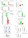
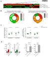
Figure 1
Figure 1. CD5L is a candidate regulator of Th17 cell functional states
(A–C) Single-cell RNA-seq analysis. (A) Cd5l expression of single-cells from in-vitro generated and in-vivo sorted Th17 cells (IL-17.GFP+) from mice at the peak of EAE. (B,C) Correlation of Cd5l expression in non-pathogenic Th17 cells (TGF-β1+IL-6) with (B) the cell pathogenicity score (based on the pathogenic signature of (Lee et al., 2012)). p = 2.63 ×10⁻⁵ (Wilcoxon ranksum test, comparing signature scores of Cd5l expressing vs. non-expressing cells); (C) the founding signature genes of the single-cell based proinflammatory (red) and regulatory (green) modules (Solid bars, significant correlation (p < 0.05); striked bars, none significant correlation). (D–F) Validation of CD5L expression in vitro. Naïve T cells (CD4+CD62L+CD44CD25) were sorted and differentiated as indicated and analyzed by qPCR for CD5L expression at 48h (D) and 72h (E) and by flow cytometry at 48h (F); (E) IL-23 or control was added at 48h in fresh media. (G–I) Validation of Cd5l expression in vivo. (G,H) IL-17A.GFP reporter mice were immunized to induce EAE. Cells were sorted from spleen (G) and CNS (H) at the peak of disease. Cd5l and Il17a expression are measured by qPCR. Figure shown is representative data of three technical replicates from two independent experiments. (I) Cells were sorted from the gut of naïve mice and the number of RNA transcripts measured by nanostring nCounter platform (supp. Experimental Procedures). See also Figure S1.
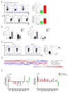
Figure 2
Figure 2. CD5L represses effector functions without affecting Th17 cell differentiation
(A) EAE was induced by MOG/CFA (40μg) immunization. Left panel is pooled results from 3 independent experiments. Right panel: cytokine profile of CD4 T cells isolated from CNS at day 15 post immunization with summary data in Figure S2B. (B–D) Naïve splenic T cells were sorted and differentiated with TGF-β1+IL-6 for 48h. Th17 cell signature genes were measured by flow cytometry (B), ELISA (C) and qPCR (D). (E–F) Effector Th17 cells were differentiated as in B and resuspended in fresh media with no cytokines for 72h followed by restimulation. Gene profile was measured by flow cytometry (E) and qPCR (F). (G–H) Effector memory T cells (CD4+CD62LCD44+) (G) or Effector memory Th17 cells (CD4+CD62LCD44+RorγtGFP+) (H) were sorted from spleen of naïve mice and activated with TCR stimulation. See also Fig S2.
Figure 3
Figure 3. CD5L is a switch that regulates Th17 cell pathogenicity
(A–D) Passive EAE is induced as previously described (Jager et al., 2009). Naïve WT or CD5L−/− 2D2 splenic T cells were differentiated with TGF-β1+IL-6 and transferred into syngeneic WT hosts. (A) Cytokine profile of 2D2 T cells after differentiation at day 4. (B) Weight and clinical score of recipient mice undergoing EAE. (C) Representative histology of optic nerve (upper 2 panels, Hematoxylin and eosin stain) and spinal cord (lower 4 panels, Luxol fast blue-hematoxylin and eosin stains). Demyelination is indicated by loss of normal blue staining of myelin in lower panels. (D) Cytokine profile of WT and CD5L−/− 2D2 cells isolated from CNS at day 27 post transfer. Cells were gated on Va3.2+CD4+. (E) CD45.1 WT recipients received 100,000 naïve WT or CD5L−/− 2D2 T cells and were immunized the following day with MOG/CFA without pertussis toxin. Cytokine profile of 2D2 T cells was examined on day 10 in draining LN. (F) Expression profile of pathogenicity signature genes in WT and CD5L−/− Th17 cells differentiated with TGF-β1+IL-6 as in Figure 2E. Data are summary of at least three independent experiments. See also Figure S3.
Figure 4
Figure 4. CD5L shifts the fatty acid composition of Th17 cell lipidome and modulates Rorγt ligand availability
(A–C) Lipidome analysis of Th17 cells showing altered SFA/MUFA and PUFA composition between WT and CD5L−/− Th17 cells (Exprimental Procedures). (A) Heatmap of 39 metabolites (rows) with significantly different levels among any Th17 cell conditions (columns) and with a fold change of at least 1.5. Median Intensity of each metabolite is shown (color bar is normalized per row). (B) Lipids from the two clusters in (A) are partitioned based on the length and saturation of their fatty acyl (FA) side chains. Those carrying more than one FA are further grouped by their FAs with the least saturation or longest carbon chain (in that order). Complete FA profile is shown in Figure S4BC. (C) Ratio of specific lipids in WT vs. CD5L−/− Th17 cells carrying various PUFA side chains. Phospholipids included in this analysis: phosphatidylcholine, phosphatidylethanolamine, phosphatidylserine and their respective lyso-metabolites. Neutral lipid included in this analysis: Triacylglyceride, diacylglyceride and monoacylglyceride. Asterisk (*) denotes to p < 0.05 in Student’s t-test. (D) Expression of cyp51 and sc4mol mRNA in WT or CD5L−/− Th17 cells (TGF-β1+IL-6, left panels) or WT Th17 cells (TGF-β1+IL-6 with control or IL-23, right panels). SFA (palmitic acid, 25uM) or PUFA (arachidonic acid, 25uM) was added at 48h and cells analyzed at 96h. See also Figure S4 and Table S1.
Figure 5
Figure 5. CD5L and PUFA/SFA profile regulate Rorγt function in a ligand-dependent manner
(A, B) Rorγt ChIP-PCR analyses in WT and CD5L−/− Th17 cells. WT, CD5L−/− and Rorγt−/− Th17 cells were differentiated with TGF-β1+IL-6 for 96h. Enrichment of Rorγt binding to genomic regions of Il17 (A) and Il10 (B) is measured using qPCR. For fatty acid experiments, 10μM of either SFA (palmitic acid) or PUFA (arachidonic acid or docosahexaenoic acid showed similar results) was added to WT Th17 cell culture at day 0. Three independent experiments were performed. (C, D) Rorγt transcriptional activity was measured by luciferase reporter of Il17 promoter in EL4 cells transfected with CD5L-RV at 0, 25, 50, 100ng (C) or 100ng with 7, 27 dihydroxycholsterol (5, 0.5 or 0.05uM) (D). (E) Naïve WT T cells were activated without polarizing cytokines (Th0) and infected with retrovirus expressing Rorγt in the presence of control-RV or CD5L-RV with or without FF-MAS (5uM) as a source of Rorγt ligand. Each dot represents an independent infection. (F) WT or CD5L−/− naïve cells were differentiated with TGF-β1+IL-6. At 48h, cells were replated in fresh media with either control or FF-MAS (5uM) as a source of Rorγt ligand. Cells were harvested for FACS analysis 72h later. See also Figure S5.
Figure 6
Figure 6. PUFA/SFA regulate Th17 cell function and contribute to CD5L-dependent regulation of Th17 cells
(A, B) Naïve T cells were sorted from WT or IL-23RGFP reporter mice (A); or WT or Rorc−/− mice (B) and differentiated with TGF-β1+IL-6 followed by addition of IL-23 at 48h in fresh media with either PUFA (arachidonic acid, 10uM) or SFA (palmitic acid, 20uM). Cells were analyzed by flow cytometry (A) or qPCR (B) at 96h. The concentration of free fatty acids was predetermined in titration experiments (data not shown). (C) Naïve WT and CD5L−/− T cells were differentiated with TGF-β1+IL-6 and replated in fresh media with control or 5uM of PUFA at 48h. Cells were analyzed at 96h. (D, E) Same as in C. PUFA (5uM) or SFA (25uM) were added 48h. Cells were analyzed by nanostring analysis (D) or qPCR (E) at 96h. (D) Genes that are differentially expressed between any of the four samples and are with a fold change of at least 1.5 are plotted in heatmap. Color scale is normalized per row (subtracted by mean and divided by standard deviation). All data are summary of 3 independent experiments. See also Table S2.

Comment in

  • T cells: Seq-ing out the 'bad' guys.
    Bird L. Bird L. Nat Rev Immunol. 2016 Jan;16(1):3. doi: 10.1038/nri.2015.15. Epub 2015 Dec 21. Nat Rev Immunol. 2016. PMID: 26688347 No abstract available.

References

    1. Ahmed M, Gaffen SL. IL-17 in obesity and adipogenesis. Cytokine & growth factor reviews. 2010;21:449–453. - PMC - PubMed
    1. Amit I, Citri A, Shay T, Lu Y, Katz M, Zhang F, Tarcic G, Siwak D, Lahad J, Jacob-Hirsch J, et al. A module of negative feedback regulators defines growth factor signaling. Nature genetics. 2007;39:503–512. - PubMed
    1. Amit I, Garber M, Chevrier N, Leite AP, Donner Y, Eisenhaure T, Guttman M, Grenier JK, Li W, Zuk O, et al. Unbiased reconstruction of a mammalian transcriptional network mediating pathogen responses. Science. 2009;326:257–263. - PMC - PubMed
    1. Arpaia N, Campbell C, Fan X, Dikiy S, van der Veeken J, deRoos P, Liu H, Cross JR, Pfeffer K, Coffer PJ, et al. Metabolites produced by commensal bacteria promote peripheral regulatory T-cell generation. Nature. 2013;504:451–455. - PMC - PubMed
    1. Awasthi A, Riol-Blanco L, Jager A, Korn T, Pot C, Galileos G, Bettelli E, Kuchroo VK, Oukka M. Cutting edge: IL-23 receptor gfp reporter mice reveal distinct populations of IL-17-producing cells. Journal of immunology. 2009;182:5904–5908. - PMC - PubMed

Publication types

MeSH terms

Substances