

MR Imaging of Fetuses to Evaluate Placental Insufficiency
- PMID: 26607809
- PMCID: PMC5600058
- DOI: 10.2463/mrms.mp.2015-0051
MR Imaging of Fetuses to Evaluate Placental Insufficiency
Abstract
Purpose: To evaluate morphological and signal intensity (SI) changes of placental insufficiency on magnetic resonance imaging (MRI) and to assess morphological changes and decreased flow voids (FVs) on T2-weighted rapid acquisition with relaxation enhancement (RARE) images for diagnosing placental insufficiency.
Methods: Fifty singleton fetuses underwent MRI using a 1.5-T MR scanner. Placental thickness, area, volume, SI, amniotic fluid SI, and size of FVs between the uterus and the placenta were measured on MR images. Two radiologists reviewed T2-weighted RARE images for globular appearance of the placenta and FVs between the uterus and the placenta. Data were analyzed using t-tests, McNemar's tests, and areas under the receiver operating characteristic curve (AUCs) at 5% level of significance.
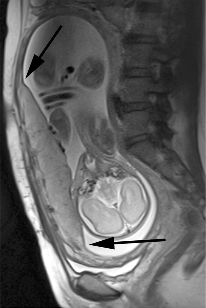
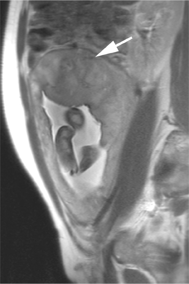
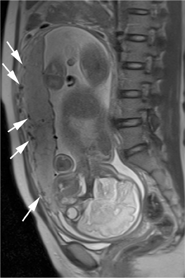
Results: Twenty-five of the 50 pregnancies were categorized as having an insufficient placenta. Significant differences were observed between insufficient and normal placentas in mean placental thickness, area, volume, placenta to amniotic fluid SI ratio, and size of FVs (49.0 mm vs. 36.9 mm, 1.62 × 10(4) mm(2) vs. 2.67 × 10(4) mm(2), 5.13 × 10(5) mm(3) vs. 6.56 × 10(5) mm(3), 0.549 vs. 0.685, and 3.4 mm vs. 4.3 mm, respectively). The sensitivity and accuracy using globular appearance plus decreased FVs were greater than those using decreased FVs (P < 0.01). There was no significant difference among AUCs using globular appearance and decreased FVs, and globular appearance plus decreased FVs.
Conclusions: Placental insufficiency was associated with placental thickness, area, volume, placenta to amniotic fluid SI ratio, and size of FVs. Evaluating FVs on T2-weighted RARE images can be useful for detecting placental insufficiency, particularly in placentas without globular appearance on MR images.
Figures
References
-
- Kaufmann P, Black S, Huppertz B. Endovascular trophoblast invasion: implications for the pathogenesis of intrauterine growth retardation and preeclampsia. Biol Reprod 2003; 69:1–7. - PubMed
-
- Lausman A, Kingdom J, Maternal Fetal Medicine Committee et al. Intrauterine growth restriction: screening, diagnosis, and management. J Obstet Gynaecol Can 2013; 35:741–757. - PubMed
-
- Sarmandal P, Grant JM. Effectiveness of ultrasound determination of fetal abdominal circumference and fetal ponderal index in the diagnosis of asymmetrical growth retardation. Br J Obstet Gynaecol 1990; 97:118–123. - PubMed
-
- Derwig I, Lythgoe DJ, Barker GJ, et al. Association of placental perfusion, as assessed by magnetic resonance imaging and uterine artery Doppler ultrasound, and its relationship to pregnancy outcome. Placenta 2013; 34:885–891. - PubMed
-
- Javor D, Nasel C, Schweim T, Dekan S, Chalubinski K, Prayer D. In vivo assessment of putative functional placental tissue volume in placental intrauterine growth restriction (IUGR) in human fetuses using diffusion tensor magnetic resonance imaging. Placenta 2013; 34:676–680. - PubMed
MeSH terms
LinkOut - more resources
Full Text Sources
Other Literature Sources
Medical