. 2016 Feb:276:5-12.
doi: 10.1016/j.expneurol.2015.11.006. Epub 2015 Nov 29.

Pycnogenol protects CA3-CA1 synaptic function in a rat model of traumatic brain injury

Affiliations

Pycnogenol protects CA3-CA1 synaptic function in a rat model of traumatic brain injury

Christopher M Norris et al. Exp Neurol. 2016 Feb.

Abstract

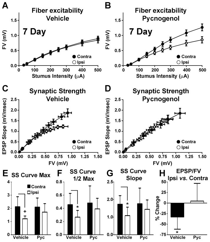
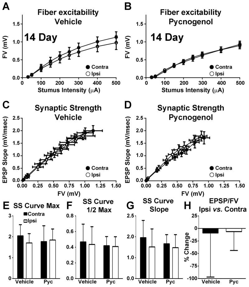
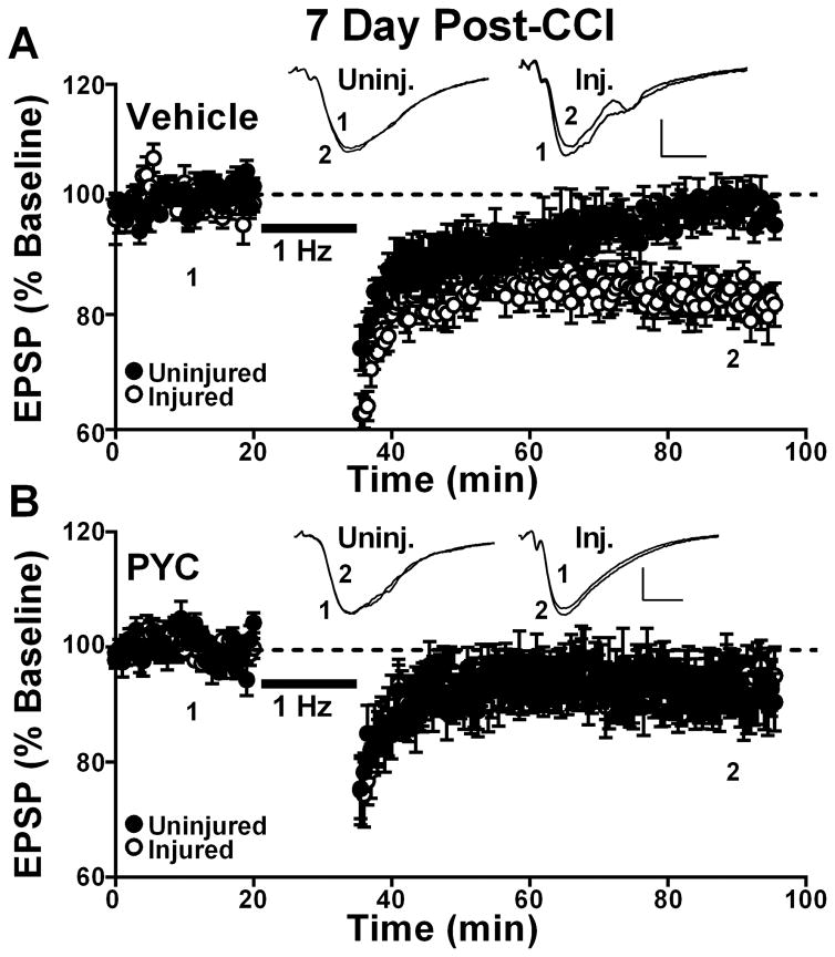
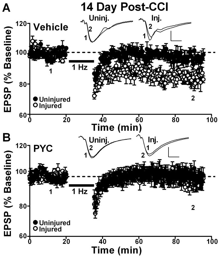
Pycnogenol (PYC) is a patented mix of bioflavonoids with potent anti-oxidant and anti-inflammatory properties. Previously, we showed that PYC administration to rats within hours after a controlled cortical impact (CCI) injury significantly protects against the loss of several synaptic proteins in the hippocampus. Here, we investigated the effects of PYC on CA3-CA1 synaptic function following CCI. Adult Sprague-Dawley rats received an ipsilateral CCI injury followed 15 min later by intravenous injection of saline vehicle or PYC (10 mg/kg). Hippocampal slices from the injured (ipsilateral) and uninjured (contralateral) hemispheres were prepared at seven and fourteen days post-CCI for electrophysiological analyses of CA3-CA1 synaptic function and induction of long-term depression (LTD). Basal synaptic strength was impaired in slices from the ipsilateral, relative to the contralateral, hemisphere at seven days post-CCI and susceptibility to LTD was enhanced in the ipsilateral hemisphere at both post-injury timepoints. No interhemispheric differences in basal synaptic strength or LTD induction were observed in rats treated with PYC. The results show that PYC preserves synaptic function after CCI and provides further rationale for investigating the use of PYC as a therapeutic in humans suffering from neurotrauma.

Keywords: Hippocampus; Oxidative stress; Pycnogenol®; Synaptic transmission; Traumatic brain injury.


Figures

Figure 1
Figure 1. Effects of PYC on CA3 axon excitability and CA3-CA1 basal synaptic strength at 7 days post CCI
(A–B) Input/output curves for mean ± SEM FV amplitudes in CA1 stratum radiatum of hippocampal slices from contralateral (Contra) and ipsilateral (Ipsi) hemispheres of rats treated with vehicle (A, n = 8 rats/group) or PYC (B, n = 9 rats). (C–D) Mean ± SEM EPSP slope measures plotted against FV measures from the same rats shown in A and B. (E–G) Mean ± SD values for synaptic strength (SS) curve parameters shown in C and D: maximum distribution amplitude (E, Max), half maximum activation level (F, 1/2 Max), and distribution slope (G, Slope). (H) Mean ± SD maximum EPSP/FV ratio. +p < 0.01, * p < 0.05 determined using repeated measures ANOVA and post hoc analyses.
Figure 2
Figure 2. Effects of PYC on CA3 axon excitability and CA3-CA1 basal synaptic strength at 7 days post CCI
(A–B) Input/output curves for mean ± SEM FV amplitudes in CA1 stratum radiatum of hippocampal slices from contralateral (Contra) and ipsilateral (Ipsi) hemispheres of rats treated with vehicle (A, n = 7 rats/group) or PYC (B, n = 8 rats/group). (C–D) Mean ± SEM EPSP slope measures plotted against FV measures from the same rats shown in A and B. (E–G) Mean ± SD values for synaptic strength (SS) curve parameters shown in C and D: maximum distribution amplitude (E, Max), half maximum activation level (F, 1/2 Max), and distribution slope (G, Slope). (H) Mean ± SD maximum EPSP/FV ratio.
Figure 3
Figure 3. Effects of PYC on LTD induction in hippocampal CA1 at 7 days post CCI
Time plots showing mean ± SEM EPSP slope measures collected in CA1 stratum radiatum of uninjured and injured hemispheres before (1) and 60 min after (2) the delivery of a 900 pulse train (1 Hz, bar) to the Schaffer collaterals. Responses are normalized to the pre-1 Hz baseline. Vehicle treated rats (n = 8) are shown in (A) and rats treated with PYC (n = 8) are shown in (B). Insets: representative field potentials averaged over 10 min before (1) and at 60 min after (2) the delivery of 1Hz stimulation. Calibration bars 0.5 mV/5 ms.
Figure 4
Figure 4. Effects of PYC on LTD induction in hippocampal CA1 at 14 days post CCI
Time plots showing mean ± SEM EPSP slope measures collected in CA1 stratum radiatum of uninjured and injured hemispheres before (1) and 60 min after (2) the delivery of a 900 pulse train (1 Hz, bar) to the Schaffer collaterals. Responses are normalized to the pre-1 Hz baseline. Vehicle treated rats (n = 7) are shown in (A) and rats treated with PYC (n = 6) are shown in (B). Insets: representative field potentials averaged over 10 min before (1) and at 60 min after (2) the delivery of 1Hz stimulation. Calibration bars 0.5 mV/5 ms.

References

    1. Agrawal R, Noble E, Tyagi E, Zhuang Y, Ying Z, Gomez-Pinilla F. Flavonoid derivative 7,8-DHF attenuates TBI pathology via TrkB activation. Biochim Biophys Acta. 2015;1852:862–872. - PMC - PubMed
    1. Albensi BC, Sullivan PG, Thompson MB, Scheff SW, Mattson MP. Cyclosporin ameliorates traumatic brain-injury-induced alterations of hippocampal synaptic plasticity. Exp Neurol. 2000;162:385–389. - PubMed
    1. Ansari MA, Keller JN, Scheff SW. Protective effect of Pycnogenol in human neuroblastoma SH-SY5Y cells following acrolein-induced cytotoxicity. Free Radic Biol Med. 2008a;45:1510–1519. - PMC - PubMed
    1. Ansari MA, Roberts KN, Scheff SW. Oxidative stress and modification of synaptic proteins in hippocampus after traumatic brain injury. Free Radic Biol Med. 2008b;45:443–452. - PMC - PubMed
    1. Ansari MA, Roberts KN, Scheff SW. Dose- and Time-Dependent Neuroprotective Effects of Pycnogenol (R) following Traumatic Brain Injury. J Neurotrauma. 2013;30:1542–1549. - PMC - PubMed

Publication types

MeSH terms