. 2015 Nov 25;90(4):2112-8.
doi: 10.1128/JVI.02678-15. Print 2016 Feb 15.

Structure of an HIV-2 gp120 in Complex with CD4

Affiliations

Structure of an HIV-2 gp120 in Complex with CD4

Yunji W Davenport et al. J Virol. .

Abstract

HIV-2 is a nonpandemic form of the virus causing AIDS, and the majority of HIV-2-infected patients exhibit long-term nonprogression. The HIV-1 and HIV-2 envelope glycoproteins, the sole targets of neutralizing antibodies, share 30 to 40% identity. As a first step in understanding the reduced pathogenicity of HIV-2, we solved a 3.0-Å structure of an HIV-2 gp120 bound to the host receptor CD4, which reveals structural similarity to HIV-1 gp120 despite divergence in amino acid sequence.


Figures

FIG 1
FIG 1
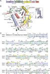
Structure and sequence of HIV-2 gp120 core. (A) Diagram of the domain structure of the gp120 core, with locations of deleted loops indicated. Branch symbols mark potential glycosylation sites. (B) Structure of the HIV-2 gp120 core, with polypeptide chains as ribbons, alpha helices as cylinders, and carbohydrates as stick models. Disordered residues are shown as dashed lines. Domains are colored as in panel A. (Inset) Annealed omit electron density map in the vicinity of the N-glycan attached to N295gp120-ST contoured at 1.5σ. (C) Alignment of the gp120 core sequences of HIV-2 and HIV-1 HxB2. Residues in blue are identical; those in green are conservative substitutions (scoring > 0.5 in the Gonnet PAM 250 matrix [49]). The mutation to remove a PNGS in HIV-2 is in red. Colors of secondary structure elements correspond to colors in panels A and B. PNGS are marked as in panel A; sites with ordered sugars are indicated with black branch symbols, while sites with disordered sugars are indicated with gray branch symbols.
FIG 2
FIG 2
Superposition of HIV-2, HIV-1, and SIV gp120 structures. (A) (Left) Sequence-based alignment of HIV-2ST gp120 core (green) with liganded and unliganded HIV-1 gp120 cores (gray) shows structural similarity. (Right) The SIVmac32H gp120 core from same viewpoint shows several secondary structural elements with different conformations. (B and C) Alignment of loops D and E from HIV-1 (various colors) and HIV-2 (mint green). (D) Alignment of the gp120 core sequences of HIV-2 and SIV. Residues in blue are identical; those in green are conservative substitutions (scoring >0.5 in the Gonnet PAM 250 matrix [49]). Colors of secondary structure elements correspond to colors in Fig. 1A. PNGSs are marked as in Fig. 1C; sites with ordered sugars are indicated with black branch symbols, while sites with disordered sugars are indicated with gray branch symbols.
FIG 3
FIG 3
Conservation of the CD4 binding site. (A) Superposition of HIV-2ST gp120 core in complex with sCD4 (green) with analogous HIV-1 cocrystal structures (gray). (B and C) Polar contacts within the gp120ST/CD4 (left) and gp120HIV-1/CD4 (right) binding interface. (D) Superposition of CD4-binding loops from HIV-2 and HIV-1 gp120 core/CD4 complex structures (colors are as in Fig. 2B).
FIG 4
FIG 4
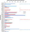
Buried per-residue accessible surface area of CD4 upon binding to HIV-2 (blue) or HIV-1 (pink) gp120. Although much of the buried interface is shared, there are residues that are contacted by only HIV-1 or HIV-2 gp120. The CCP4 program AREAIMOL (mode DIFFMODE COMPARE) was used to calculate the per-residue difference in accessible surface area (1.4-Å probe radius) for CD4 upon complex formation with Env. HIV-1 coordinates were from PDB entry 1RZJ.

References

    1. Kong R, Li H, Georgiev I, Changela A, Bibollet-Ruche F, Decker JM, Rowland-Jones SL, Jaye A, Guan Y, Lewis GK, Langedijk JP, Hahn BH, Kwong PD, Robinson JE, Shaw GM. 2012. Epitope mapping of broadly neutralizing HIV-2 human monoclonal antibodies. J Virol 86:12115–12128. doi:10.1128/JVI.01632-12. - DOI - PMC - PubMed
    1. Kong R, Li H, Bibollet-Ruche F, Decker JM, Zheng NN, Gottlieb GS, Kiviat NB, Sow PS, Georgiev I, Hahn BH, Kwong PD, Robinson JE, Shaw GM. 2012. Broad and potent neutralizing antibody responses elicited in natural HIV-2 infection. J Virol 86:947–960. doi:10.1128/JVI.06155-11. - DOI - PMC - PubMed
    1. de Silva TI, Cotten M, Rowland-Jones SL. 2008. HIV-2: the forgotten AIDS virus. Trends Microbiol 16:588–595. doi:10.1016/j.tim.2008.09.003. - DOI - PubMed
    1. Hahn BH, Kong LI, Lee SW, Kumar P, Taylor ME, Arya SK, Shaw GM. 1987. Relation of HTLV-4 to simian and human immunodeficiency-associated viruses. Nature 330:184–186. doi:10.1038/330184a0. - DOI - PubMed
    1. van der Loeff MF, Larke N, Kaye S, Berry N, Ariyoshi K, Alabi A, van Tienen C, Leligdowicz A, Sarge-Njie R, da Silva Z, Jaye A, Ricard D, Vincent T, Jones SR, Aaby P, Jaffar S, Whittle H. 2010. Undetectable plasma viral load predicts normal survival in HIV-2-infected people in a West African village. Retrovirology 7:46. doi:10.1186/1742-4690-7-46. - DOI - PMC - PubMed

Publication types