

Nicotinic acetylcholine receptor expression in human airway correlates with lung function
- PMID: 26608528
- PMCID: PMC4888556
- DOI: 10.1152/ajplung.00101.2015
Nicotinic acetylcholine receptor expression in human airway correlates with lung function
Abstract
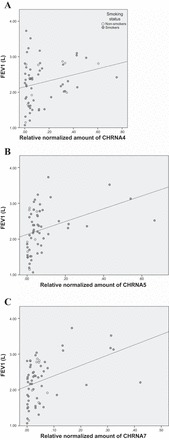
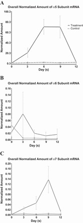
Nicotine and its derivatives, by binding to nicotinic acetylcholine receptors (nAChRs) on bronchial epithelial cells, can regulate cellular signaling and inflammatory processes. Delineation of nAChR subtypes and their responses to nicotine stimulation in bronchial epithelium may provide information for therapeutic targeting in smoking-related inflammation in the airway. Expression of nAChR subunit genes in 60 bronchial epithelial biopsies and immunohistochemical staining for the subcellular locations of nAChR subunit expression were evaluated. Seven human bronchial epithelial cell lines (HBECs) were exposed to nicotine in vitro for their response in nAChR subunit gene expression to nicotine exposure and removal. The relative normalized amount of expression of nAChR α4, α5, and α7 and immunohistochemical staining intensity of nAChR α4, α5, and β3 expression showed significant correlation with lung function parameters. Nicotine stimulation in HBECs resulted in transient increase in the levels of nAChR α5 and α6 but more sustained increase in nAChR α7 expression. nAChR expression in bronchial epithelium was found to correlate with lung function. Nicotine exposure in HBECs resulted in both short and longer term responses in nAChR subunit gene expression. These results gave insight into the potential of targeting nAChRs for therapy in smoking-related inflammation in the airway.
Keywords: bronchial epithelium; lung function; nicotine; nicotinic acetylcholine receptor; quantitative polymerase chain reaction.
Copyright © 2016 the American Physiological Society.
Figures
References
-
- Colombo SF, Mazzo F, Pistillo F, Gotti C. Biogenesis, trafficking and up-regulation of nicotinic ACh receptors. Biochem Pharmacol 86: 1063–1073, 2013. - PubMed
-
- De Luca V, Likhodi O, Van Tol HH, Kennedy JL, Wong AH. Regulation of alpha7-nicotinic receptor subunit and alpha7-like gene expression in the prefrontal cortex of patients with bipolar disorder and schizophrenia. Acta Psychiatr Scand 114: 211–215, 2006. - PubMed
-
- Gerzanich V, Wang F, Kuryatov A, Lindstrom J. α5 Subunit alters desensitization, pharmacology, Ca++ permeability and Ca++ modulation of human neuronal alpha 3 nicotinic receptors. J Pharmacol Exp Ther 286: 311–320, 1998. - PubMed
-
- Global Initiative for Chronic Obstructive Lung Disease. Global Strategy for the Diagnosis, Management, and Prevention of Chronic Obstructive Pulmonary Disease. Available from http://www.goldcopd.org, 2015. - PubMed
Publication types
MeSH terms
Substances
Grants and funding
LinkOut - more resources
Full Text Sources
Other Literature Sources