. 2015 Nov 26:5:17189.
doi: 10.1038/srep17189.

Matrix metalloproteinase-12 is an essential mediator of acute and chronic arterial stiffening

Affiliations

Matrix metalloproteinase-12 is an essential mediator of acute and chronic arterial stiffening

Shu-Lin Liu et al. Sci Rep. .

Abstract

Arterial stiffening is a hallmark of aging and risk factor for cardiovascular disease, yet its regulation is poorly understood. Here we use mouse modeling to show that matrix metalloproteinase-12 (MMP12), a potent elastase, is essential for acute and chronic arterial stiffening. MMP12 was induced in arterial smooth muscle cells (SMCs) after acute vascular injury. As determined by genome-wide analysis, the magnitude of its gene induction exceeded that of all other MMPs as well as those of the fibrillar collagens and lysyl oxidases, other common regulators of tissue stiffness. A preferential induction of SMC MMP12, without comparable effect on collagen abundance or structure, was also seen during chronic arterial stiffening with age. In both settings, deletion of MMP12 reduced elastin degradation and blocked arterial stiffening as assessed by atomic force microscopy and immunostaining for stiffness-regulated molecular markers. Isolated MMP12-null SMCs sense extracellular stiffness normally, indicating that MMP12 causes arterial stiffening by remodeling the SMC microenvironment rather than affecting the mechanoresponsiveness of the cells themselves. In human aortic samples, MMP12 levels strongly correlate with markers of SMC stiffness. We conclude that MMP12 causes arterial stiffening in mice and suggest that it functions similarly in humans.


Figures
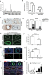
Figure 1
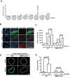
Figure 1. Preferential induction of MMP12 is causal for arterial stiffening after vascular injury.
Male wild-type and MMP12-null mice were subjected to femoral artery injury. (A) Differential expression of MMP mRNAs after femoral artery injury in 4–5 month old wild-type mice. (B) Uninjured (n = 6) and injured (n = 4) femoral arteries from wild-type and MMP12-null mice were isolated, cleaned, and immediately analyzed by AFM. The bar graph shows mean + SE. (C) Cyclin D1 staining of uninjured and injured femoral artery sections. Dashed lines show the internal elastic lamina (IEL) and external elastic laminae (EEL). M; media. NI; neointima. Scale bar = 50 μm. (D) Blind quantification of results in C from wild-type (n = 10) and MMP12-null (n = 8) mice. Statistical significance was determined by chi-square test. (E) In situ elastase activity of cross sections of uninjured and injured femoral arteries. Scale bar = 50 μm. (F) Quantification of results in panel E from wild-type (n = 7) and MMP12-null (n = 5) mice. The bar graph shows mean + SE. (G) Elastin fragmentation in the IEL was detected by autofluorescence imaging (green). Scale bar = 25 μm. Nuclei (blue) were stained with DAPI. (H) Quantification of results in panel G from wild-type (n = 10) and MMP12-null (n = 8) mice. The bar graph shows mean + SE. (I) Cross sections of uninjured and injured femoral arteries of wild-type mice immunostained for MMP12 (red) and SMCs (anti-SMA, green) (n = 8). Nuclei (blue) were stained with DAPI. Dashed lines show the IEL and EEL as determined by autofluorescence. (J) Differentiated and de-differentiated SMCs were serum-starved and incubated with 1 mg/ml heat-inactivated, fatty acid-free BSA in the absence (control) or presence of PDGF for 24 h. MMP12 mRNA levels were determined by RT-qPCR and plotted relative to 18S rRNA. The bar graph shows mean + SD; n = 4. The levels of MMP12 mRNA in the absence of PDGF was set to 1.0. All p values are from two-tailed Mann-Whitney tests.
Figure 2
Figure 2. MMP12 mediates age-dependent arterial stiffening.
Aortas were isolated from male wild-type and MMP12-null mice of different ages. (A) The samples were analyzed by real-time qPCR. Results show mean + SD; n = 2 with each independent experiment representing a pool of 4 aortas. The levels of each mRNA in the 2-month arteries were set to 1.0. (B) Cross sections of aortic roots from 2 (n = 7), 6 (n = 8) and 12 (n = 8) month-old male wild-type mice were stained for SMA (green) and MMP12 (red). Scale bar = 50 μm. (C) AFM of aortas from 2–18 month-old wild-type (n = 4) and MMP12-null (n = 4) mice. The bar graph shows mean + SE. (D) In situ elastase activity in cross sections of aortic roots from 6 and 12 month-old wild-type and MMP12-null mice. Scale bar = 50 μm. (E) Quantification of results in panel D from wild-type (n = 6) and MMP12-null (n = 8) mice. The bar graph shows mean + SE. All p values are from two-tailed Mann-Whitney tests.
Figure 3
Figure 3. Similar effects of ECM stiffness on wild-type and MMP12-null SMCs in culture.
(A) Mouse (n = 4) and human (n = 5) vascular SMCs were serum-starved for 48 h and then seeded on low and high stiffness fibronectin-coated acrylamide hydrogels with 10% FBS for 24 h. The bar graph shows mean + SD with the level of MMP12 mRNA in the low stiffness hydrogels set to 1.0. (B–D) Wild-type and MMP12-null SMCs were serum-starved and incubated on low (L)-, medium (M), or high- (H)-stiffness hydrogels (2–4, 10–12, and 20–25 kPa, respectively) with 10% FBS for 24 hr. (B) Intracellular stiffness was determined by AFM. The bar graph shows mean + SE of 4 independent experiments with 10 cells analyzed per experiment. (C) Cells were co-stained with phalloidin (red) and anti-paxillin (green). Replicate coverslips were stained with anti-FAKPY397 (red); n = 4 independent experiments with 5 cells analyzed per experiment. (D) S-phase entry was determined by EdU incorporation after a 72-hr incubation in 10% FBS. The bar graph shows mean + SD; n = 4. p values are from two-tailed Mann-Whitney tests.
Figure 4
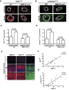
Figure 4. MMP12 expression predicts SMC stiffness.
(A,B) Cross sections of uninjured and injured femoral arteries of male wild-type [uninjured (n = 8) and injured (n = 10)] and MMP12-null [uninjured (n = 8) and injured (n = 8)] mice were immunostained for FAKpY397 and p130CaspY410. Dashed lines show the internal and external elastic laminae as determined by autofluorescence. M; media. NI; neointima. Scale bar = 50 μm. (C,D) Quantification of results from panels (A,B). The bar graph shows mean + SE. (E) Single cross sections of human ascending aortas (n = 12) immunostained for MMP12, FAKpY397, or p130CaspY410. Results from three different patients with low (L), medium (M), and high (H) MMP12 staining are shown. (F,G) Linear regression analysis of all patients (n = 12) after immunostaining for MMP12 and either FAKpY397 or p130CaspY410.

References

    1. Steppan J., Barodka V., Berkowitz D. E. & Nyhan D. Vascular stiffness and increased pulse pressure in the aging cardiovascular system. Cardiol Res Pr. 2011, 263585 (2011). - PMC - PubMed
    1. Duprez D. A. & Cohn J. N. Arterial stiffness as a risk factor for coronary atherosclerosis. Curr. Atheroscler. Rep. 9, 139–144 (2007). - PubMed
    1. Mitchell G. F. et al. Arterial stiffness and cardiovascular events: the Framingham Heart Study. Circulation 121, 505–511 (2010). - PMC - PubMed
    1. Sutton-Tyrrell K. et al. Elevated aortic pulse wave velocity, a marker of arterial stiffness, predicts cardiovascular events in well-functioning older adults. Circulation 111, 3384–3390 (2005). - PubMed
    1. Van Popele N. M. et al. Association between arterial stiffness and atherosclerosis: the Rotterdam Study. Stroke 32, 454–460 (2001). - PubMed

Substances