. 2015 Nov 26:5:17191.
doi: 10.1038/srep17191.

Rescue from tau-induced neuronal dysfunction produces insoluble tau oligomers

Affiliations

Rescue from tau-induced neuronal dysfunction produces insoluble tau oligomers

Catherine M Cowan et al. Sci Rep. .

Abstract

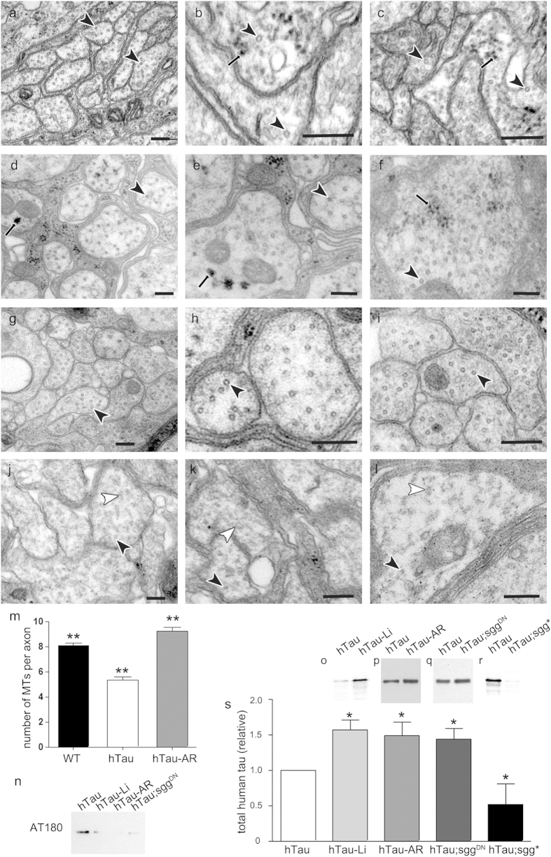
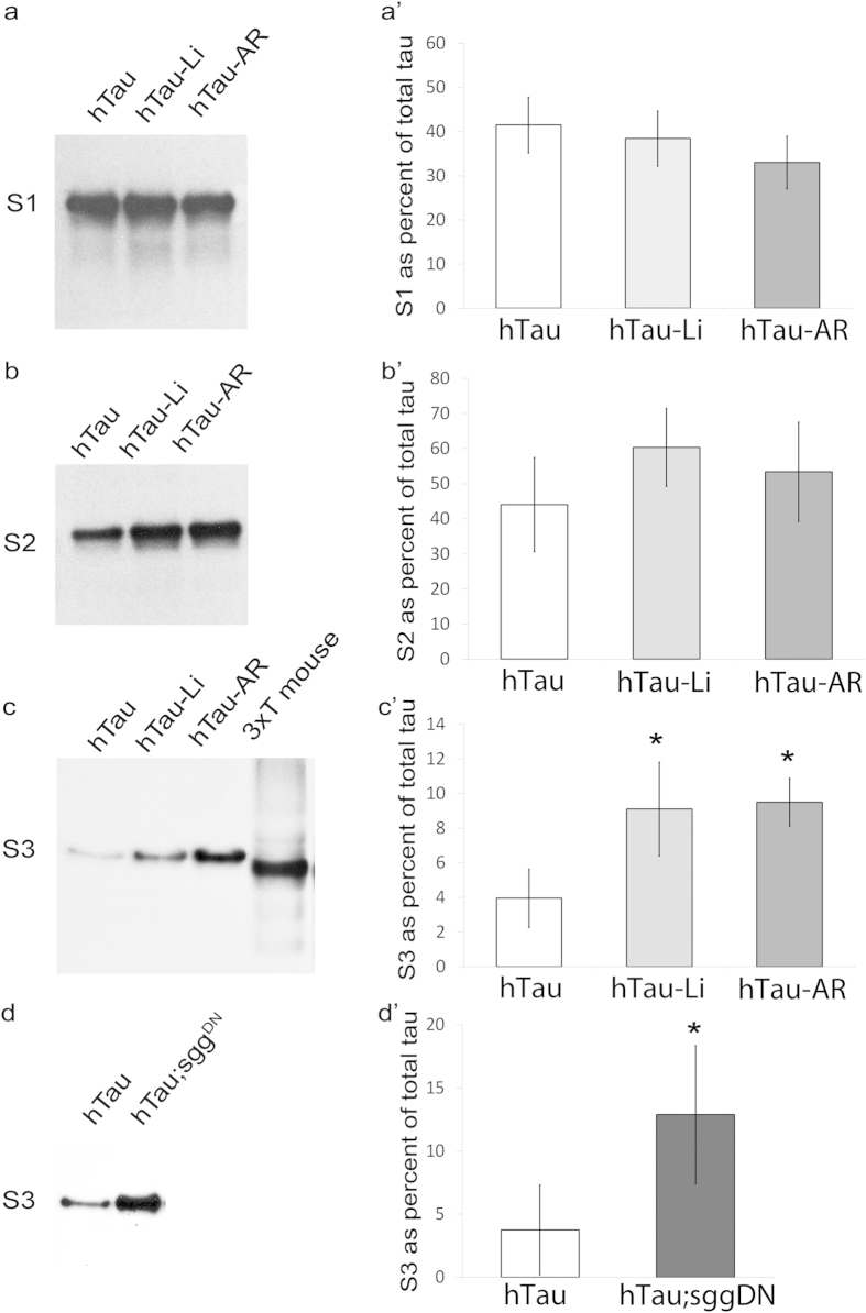
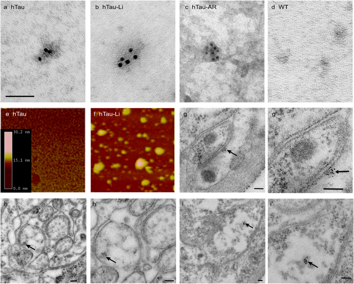
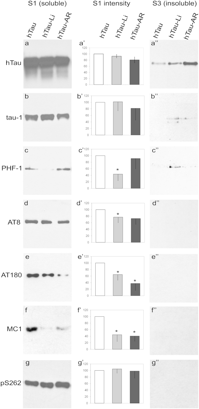
Aggregation of highly phosphorylated tau is a hallmark of Alzheimer's disease and other tauopathies. Nevertheless, animal models demonstrate that tau-mediated dysfunction/toxicity may not require large tau aggregates but instead may be caused by soluble hyper-phosphorylated tau or by small tau oligomers. Challenging this widely held view, we use multiple techniques to show that insoluble tau oligomers form in conditions where tau-mediated dysfunction is rescued in vivo. This shows that tau oligomers are not necessarily always toxic. Furthermore, their formation correlates with increased tau levels, caused intriguingly, by either pharmacological or genetic inhibition of tau kinase glycogen-synthase-kinase-3beta (GSK-3β). Moreover, contrary to common belief, these tau oligomers were neither highly phosphorylated, and nor did they contain beta-pleated sheet structure. This may explain their lack of toxicity. Our study makes the novel observation that tau also forms non-toxic insoluble oligomers in vivo in addition to toxic oligomers, which have been reported by others. Whether these are inert or actively protective remains to be established. Nevertheless, this has wide implications for emerging therapeutic strategies such as those that target dissolution of tau oligomers as they may be ineffective or even counterproductive unless they act on the relevant toxic oligomeric tau species.


Figures

Figure 1
Figure 1. GSK-3β inhibition rescued microtubule number in hTau0N3R Drosophila, but increased total hTau protein and caused formation of electron-dense granules a-l) Electron micrographs of transverse sections of peripheral nerves in L3 Drosophila (scale bar 200 nm).
In hTau-expressing (elavC155-Gal4/Y; UAS-hTau0N3R/+) animals treated with either 20 mM LiCl (hTau-Li, a–c) or with 20 μM AR-A01448 (hTau-AR, d-f), some axons exhibited small electron-dense globular structures of approximately 20–50 nm in size (black arrows). These structures were extremely rare in control larvae expressing elavC155-Gal4 driver alone (WT, gi) or untreated hTau0N3R-expressing neurons (jl). In WT larvae the axon profiles showed numerous regularly-spaced, correctly-aligned transverse microtubule profiles (black arrowheads in g-i; 8.1 ± 0.2/axon profile). As we have previously shown, in hTau0N3R-expressing axons the microtubules were dramatically disrupted, with fewer correctly-aligned transverse microtubule profiles (black arrowheads in j-l; 5.3 ± 0.3/axon profile), and evidence of disorganised microtubules in the same axon profiles (white arrowheads in jl). Indeed, approximately 30% of hTau0N3R-expressing axons displayed no visible microtubule profiles (Figure S1). In hTau0N3R-expressing larvae fed with Li (ac) or AR (df), there were significantly more correctly-aligned transverse microtubule profiles (black arrowheads in a-l; 9.2 ± 0.3/axon) and fewer misaligned microtubules. Microtubule numbers per axon are quantified in m (**p < 0.01, unpaired Students t test). Representative Western blots of hTau0N3R-expressing fly head lysates showed that tau phosphorylation was decreased (at T231/S235 detected by AT180) whilst total tau levels were increased by 40–60% (or) by 20 mM lithium treatment (hTau-Li, o), 20 μM AR-A01448 treatment (hTau-AR, p), co-expression of dominant negative shaggy (hTau;sggDN, q) {elavC155-Gal4/Y; UAS- hTau0N3R/+ ; UAS-sggDN/+}. Conversely, total tau levels were decreased by approximately 50% by co-expression of constitutively active shaggy (hTau;sgg*, r) {elavC155-Gal4/Y; UAS- hTau0N3R/+ ; UAS-sgg*/+}. This is quantified in s (error bars are standard error of mean; *p < 0.05 by Students t-test).
Figure 2
Figure 2. The amount of insoluble tau detected from hTau0N3R-expressing Drosophila is increased dramatically after treatment with GSK-3β inhibitors.
Western blots of aqueous-soluble fraction (S1), detergent-soluble fraction (S2) and insoluble fraction (S3) probed for anti-hTau. Samples in lanes 1–3 are from heads of hTau0N3R flies (hTau – {elavC155-Gal4/Y ; UAS-hTau0N3R/+}), hTau0N3R flies treated with 20 mM lithium (hTau-Li), hTau0N3R flies treated with 20 μM AR-A01448 (hTau-AR), and flies co-expressing hTau0N3R with dominant negative shaggy (hTau;sggDN {elavC155-Gal4/Y; UAS- hTau0N3R/+; UAS-sggDN/+}). The fourth lane in the third panel (labelled “3xT mouse”) is a 10-fold dilution of sample from triple-transgenic mouse, used as a positive control for insoluble tau. Bar charts in a’–d’ are quantifications of blots in a – d presented as a percentage of total tau {S3/(S1 + S2 + S3)} in each genotype. Treatment with Li or AR-AR01448 did not alter the amount of tau detected in either the S1 (a,a’) or S2 fractions (b,b’). However treatment with Li, AR-AR01448 (c,c’) or co-expression of dominant negative shaggy (hTau;sggDN) (d,d’) significantly increased the amount of tau detected in the insoluble S3 fraction. (error bars are standard error of mean and n = 5 for each genotype/treatment; *p < 0.05 by Students t-test).
Figure 3
Figure 3. Insoluble tau oligomers can be purified from hTau0N3R-expressing Drosophila and were increased dramatically after treatment with GSK-3β inhibitors.
TEM of immuno-gold labelling for anti-hTau of S3 insoluble fractions show granular tau oligomers comprised of hTau in all conditions expressing hTau {elavC155-Gal4/Y; UAS-hTau0N3R/+}: (a) hTau, (b) hTau-Li, (c) hTau-AR. No such structures were detected in controls: d) wild-type. (See also Supplementary Fig. 8, for additional controls of no sample labelled with anti h-Tau; and hTau labelled for an irrelevant rabbit polyclonal antibody, anti-v-glut). Scale bar in a (applicable to af) = 100 nm. (gh); hundreds of such structures were observed in preparations from 18 pooled flies) Atomic Force Microscopy of material immuno-precipiated from fly head lysates using anti-hTau antibody shows the appearance of numerous granular tau oligomers present after LiCl treatment (f) but only very sparse in untreated hTau0N3R flies (e). Oligomer sizes were determined by cross-sectional height analysis of individual oligomers. The heights of the oligomers ranged between 15 and 30 nm, with a mean height of 17.07 nm (SD = 8.86). The widths of the majority of oligomers are between 20 and 40 nm and the average width was calculated to be 20.6 nm (SD = 11.4). A minority of oligomers have a larger width than 30 nm but the height of the oligomers was consistently 30 nm or below. Scale bar in i = 1 μm. (gi)’) Immuno-gold labeling for hTau in situ in sections of peripheral nerves from hTau-Li flies demonstrates labeled granular tau oligomers (arrows) within axons. Examples are given at lower magnifications (gi) in which axonal profiles are clearer, and at higher magnifications (g’–i’) in which GTOs can be seen more clearly. Scale bars = 100 nm.
Figure 4
Figure 4. Tau within GTO-like structures is largely unphosphorylated Western blots of the soluble fraction (S1) and the insoluble fraction (S3) from hTau {elavC155-Gal4/Y; UAS- hTau0N3R/ + }, hTau-Li and hTau – AR-A01448 treated fly brain lysates probed with an antibody that detects total tau (a and a”) and those that detect various phospho-tau epitopes (b-g and b”-g”).
Though there is a significant amount of tau in the insoluble fraction of drug treated brain lysates (a”), it is largely unphosphorylated at many sites (b”–g”). Signal at these sites in the soluble fractions (bg and b’–g’) provides a positive control for the antibodies, and shows that treatment with GSK-3β inhibitors LiCl and AR-A01448 reduced phosphorylation at many of these sites: quantified in graphs (b’–g’). Graphs show the average of 6 independent experiments; error bars are SEM; * p < 0.05. Blots were probed with (a) dako polyclonal anti-tau (total tau), (b) anti-tau-1 (unphosphorylated at 192–204), (c) anti-PHF-1 (pS396, pS404), (d) anti-AT8 (pS202, pT205), (e) anti-AT180 (pT231, pS235), (f) anti-MC1 (it is curious that this supposedly conformation specific anti-body picks up its epitope after SDS-PAGE denaturation; it is likely that the epitope recognized here is largely denatured but nonetheless is disease predictive since others, like us have also shown similar WB immunoreactivity in another Drosophila model of tauopathy10), (g) anti-pS262.
Figure 5
Figure 5. Raman spectroscopy indicates oligomeric structure and lack of β-pleated sheet Raman spectroscopy was carried out on the P2 (detergent-insoluble GTO) fraction prepared from fly heads.
The spectrum of Li-treated hTau {elavC155-Gal4/Y; UAS- hTau0N3R/ + } P2 fraction (black) contained several peaks (arrows) of reduced bandwidth and greater intensity than that from untreated hTau fractions (red), indicating more oligomeric structure in hTau-Li. No peaks are observed at 1665–1690 cm−1 (arrowhead) indicating a lack of β-pleated sheet structure or at 980–1000 cm−1 indicating lack of phosphorylated species.

References

    1. Frost B., Gotz J. & Feany M. B. Connecting the dots between tau dysfunction and neurodegeneration. Trends Cell Biol. 25, 46–53, doi: 10.1016/j.tcb.2014.07.005 (2015). - DOI - PMC - PubMed
    1. Boutajangout A. & Wisniewski T. Tau-based therapeutic approaches for Alzheimer’s disease - a mini-review. Gerontol. 60, 381–385, doi: 10.1159/000358875 (2014). - DOI - PMC - PubMed
    1. Grundke-Iqbal I. et al. Abnormal phosphorylation of the microtubule-associated protein tau (tau) in Alzheimer cytoskeletal pathology. PNAS 83, 4913–4917 (1986). - PMC - PubMed
    1. Hanger D. P., Hughes K., Woodgett J. R., Brion J. P. & Anderton B. H. Glycogen synthase kinase-3 induces Alzheimer’s disease-like phosphorylation of tau: generation of paired helical filament epitopes and neuronal localisation of the kinase. Neurosci. Lett. 147, 58–62 (1992). - PubMed
    1. Lovestone S. et al. Alzheimer’s disease-like phosphorylation of the microtubule-associated protein tau by glycogen synthase kinase-3 in transfected mammalian cells. Curr. Biol. 4, 1077–1086 (1994). - PubMed

Publication types

MeSH terms