. 2016 Jan 1;196(1):39-43.
doi: 10.4049/jimmunol.1501190. Epub 2015 Nov 25.

Cutting Edge: Nonobese Diabetic Mice Deficient in Chromogranin A Are Protected from Autoimmune Diabetes

Affiliations

Cutting Edge: Nonobese Diabetic Mice Deficient in Chromogranin A Are Protected from Autoimmune Diabetes

Rocky L Baker et al. J Immunol. .

Abstract

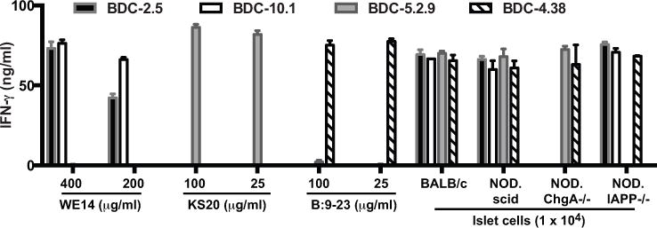
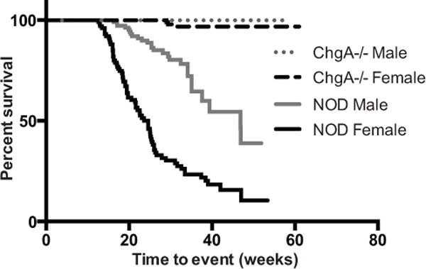
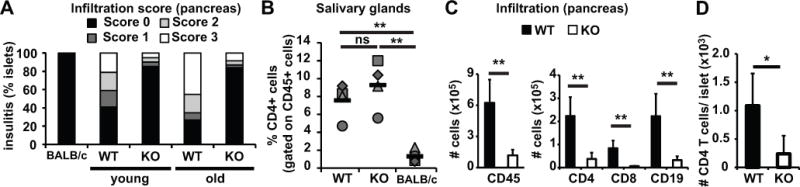
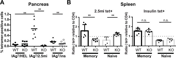
T cells reactive to β cell Ags are critical players in the development of autoimmune type 1 diabetes. Using a panel of diabetogenic CD4 T cell clones derived from the NOD mouse, we recently identified the β cell secretory granule protein, chromogranin A (ChgA), as a new autoantigen in type 1 diabetes. CD4 T cells reactive to ChgA are pathogenic and rapidly transfer diabetes into young NOD recipients. We report in this article that NOD.ChgA(-/-) mice do not develop diabetes and show little evidence of autoimmunity in the pancreatic islets. Using tetramer analysis, we demonstrate that ChgA-reactive T cells are present in these mice but remain naive. In contrast, in NOD.ChgA(+/+) mice, a majority of the ChgA-reactive T cells are Ag experienced. Our results suggest that the presence of ChgA and subsequent activation of ChgA-reactive T cells are essential for the initiation and development of autoimmune diabetes in NOD mice.


Figures

FIGURE 1
FIGURE 1. ChgA-reactive T cell clones are not activated by ChgA-deficient islets
BDC T cell clones (2 × 10⁴) were challenged with islets or peptides, in the presence of peritoneal exudate cells (2.5 × 10⁴) as antigen-presenting cells. After 24 h of culture, supernatants from duplicate wells were harvested and IFN-γ was detected by ELISA. Data is representative of two independent experiments.
FIGURE 2
FIGURE 2. ChgA-deficient NOD mice are protected from diabetes
Male or female wt NOD (female n=101; male n=110) or NOD.ChgA−/− mice (female n=118; male n=109) were followed for the development of diabetes by monitoring urine and blood glucose levels. The impact of the microbiota was not assessed and littermate controls were not monitored for disease incidence. Statistical significance was assessed by using the Wilcoxon rank-sum test.
FIGURE 3
FIGURE 3. Chromogranin A-deficient mice are protected from insulitis but not sialitis
(A) Pancreatic sections from BALB/c, NOD (WT) or NOD.ChgA−/− (KO) mice were stained with H&E and analyzed for the presence of mononuclear infiltrate in the islets. Thirty-six islets were scored for BALB/c mice. The average age for younger NOD mice was 14.6 wks (n=6, 66 islets scored) and 13.25 weeks for NOD.ChgA−/− (n=4, 61 islets scored); for older mice, the average age was 42 weeks for NOD (n=13, 119 islets scored) and 52 weeks for NOD.ChgA−/− (n=14, 275 islets scored). (B) Submaximal salivary glands from 6–10 month-old wt NOD, NOD.ChgA−/− or BALB/c mice were analyzed by flow cytometry. The percentages of CD4 cells present in submaximal salivary glands are indicated. Gates were set on the lymphocyte gate, singlets and on CD45+ and dump- (CD8- CD11b- CD11c- CD19-). Each symbol represents an individual mouse. Data is representative of two independent experiments with 2 mice per group per experiment. Averages are indicated as black horizontal bars. (C) We also investigated islet infiltration by flow cytometric analysis of cells in the pancreas. Single cell suspension from pancreata from 12–16 week-old NOD (WT) or NOD.ChgA−/− (KO) female mice were stained with anti-CD45 (BV421), anti-CD4 (FITC), anti-CD8 (APC-Cy7) and anti-CD19 (PerCP-Cy5.5) and analyzed by flow cytometry. Data are a summary of four independent experiments with 2 mice per group per experiment (n = 8). (D) Islets of wt NOD or NOD.ChgA−/− mice were hand picked, counted, dissociated into single cell suspensions and stained with appropriate antibodies. Gates were set on the lymphocyte gate, singlets, CD8-, CD4+. Data is a summary of 2 independent experiments with 2 mice per group per experiment (n = 4). Black columns represent NOD mice (WT) and white columns represent NOD.ChgA−/− mice (KO). Significance: ns: no significance (P > 0.05); ** = P < 0.01; * = P < 0.05.
FIGURE 4
FIGURE 4. ChgA-reactive T cells develop in the absence of ChgA but remain naïve
Single cell suspensions from pancreas (A) and spleen (B) were stained with tetramers specific for T cells reactive to HEL, ChgA (2.5mi), or insulin (Insp8G), and a master mix of antibodies. Gates were set on the lymphocyte gate, singlets, CD45, dump- (CD8, CD11b, CD11c, CD19, F4/80 and 7AAD) and CD4+. (A) Data shows the percentage of tetramer-positive cells in the pancreas of 12–20 week-old NOD (WT) or NOD.ChgA−/− (KO) female mice (n = 8). Each symbol represents an individual mouse and averages are indicated as a black horizontal bar. (B) Memory versus naïve cells in spleens of NOD (WT) or NOD.ChgA−/− (KO) female mice (n = 6). Data is expressed as ratios of percentages of tetramer-positive cells relative to percentages of polyclonal CD4 population, for memory vs naïve populations. Memory CD4 T cells were defined as CD44hiCD62Llo and naïve as CD44loCD62Lhi; each symbol represents an individual mouse. The dotted line indicates a ratio of 1. Data are from four (A) and three (B) independent experiments with 2 mice per group per experiment. Significance: n.s.: no significance (P > 0.05); ** = P < 0.01; * = P < 0.05.

References

    1. Stadinski BD, Delong T, Reisdorph N, Reisdorph R, Powell RL, Armstrong M, Piganelli JD, Barbour G, Bradley B, Crawford F, Marrack P, Mahata SK, Kappler JW, Haskins K. Chromogranin A is an autoantigen in type 1 diabetes. Nat Immunol. 2010;11:225–231. - PMC - PubMed
    1. Gottlieb PA, Delong T, Baker RL, Fitzgerald-Miller L, Wagner R, Cook G, Rewers MR, Michels A, Haskins K. Chromogranin A is a T cell antigen in human type 1 diabetes. J Autoimmun. 2014;50:38–41. - PMC - PubMed
    1. Delong T, Baker RL, He J, Barbour G, Bradley B, Haskins K. Diabetogenic T-cell clones recognize an altered peptide of chromogranin A. Diabetes. 2012;61:3239–3246. - PMC - PubMed
    1. Baker RL, Delong T, Barbour G, Bradley B, Nakayama M, Haskins K. Cutting edge: CD4 T cells reactive to an islet amyloid polypeptide peptide accumulate in the pancreas and contribute to disease pathogenesis in nonobese diabetic mice. J Immunol. 2013;191:3990–3994. - PMC - PubMed
    1. Yamamoto T, Yamato E, Tashiro F, Sato T, Noso S, Ikegami H, Tamura S, Yanagawa Y, Miyazaki JI. Development of autoimmune diabetes in glutamic acid decarboxylase 65 (GAD65) knockout NOD mice. Diabetologia. 2004;47:221–224. - PubMed

Publication types

MeSH terms