Review
. 2015 Nov 25;80(1):1-43.
doi: 10.1128/MMBR.00019-15. Print 2016 Mar.

Taxonomy, Physiology, and Natural Products of Actinobacteria

Affiliations
Review

Taxonomy, Physiology, and Natural Products of Actinobacteria

Essaid Ait Barka et al. Microbiol Mol Biol Rev. .

Erratum in

Abstract

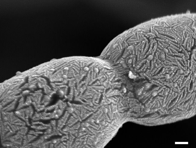
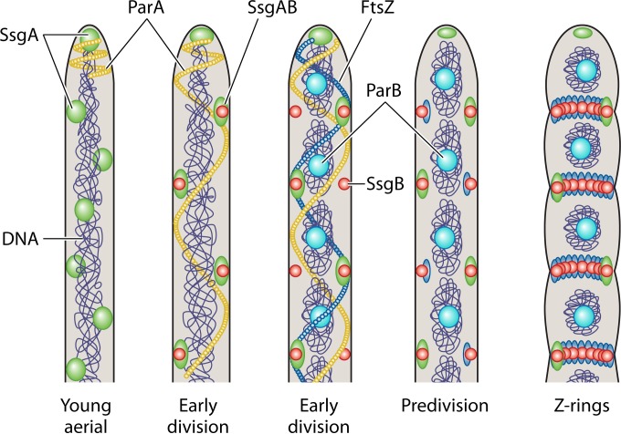
Actinobacteria are Gram-positive bacteria with high G+C DNA content that constitute one of the largest bacterial phyla, and they are ubiquitously distributed in both aquatic and terrestrial ecosystems. Many Actinobacteria have a mycelial lifestyle and undergo complex morphological differentiation. They also have an extensive secondary metabolism and produce about two-thirds of all naturally derived antibiotics in current clinical use, as well as many anticancer, anthelmintic, and antifungal compounds. Consequently, these bacteria are of major importance for biotechnology, medicine, and agriculture. Actinobacteria play diverse roles in their associations with various higher organisms, since their members have adopted different lifestyles, and the phylum includes pathogens (notably, species of Corynebacterium, Mycobacterium, Nocardia, Propionibacterium, and Tropheryma), soil inhabitants (e.g., Micromonospora and Streptomyces species), plant commensals (e.g., Frankia spp.), and gastrointestinal commensals (Bifidobacterium spp.). Actinobacteria also play an important role as symbionts and as pathogens in plant-associated microbial communities. This review presents an update on the biology of this important bacterial phylum.


Figures

FIG 1
FIG 1
Schematic representation of the life cycle of sporulating actinomycetes.
FIG 2
FIG 2
Schematic drawings of the different types of spore chains produced by actinomycetes.
FIG 3
FIG 3
A genome-based phylogenetic tree based on 97 genome sequences of the phylum Actinobacteria. Type strain genome projects were selected as previously described (676), provided that they yielded at most 25 contigs. Phylogenetic reconstruction, including the assessment of branch support, was done using amino acid sequences according to the methods described by Meier-Kolthoff et al. (677, 678). The tree was visualized by using ITOL (679). Branch support values below 60% are not shown, but the tree generally reveals high support throughout.
FIG 4
FIG 4
Major events during development of Streptomyces. Nutrient stress is a major trigger of development, leading to the accumulation of ppGpp, resulting in cessation of early growth and repression of the nutrient sensory DasR protein by cell wall-derived metabolites following PCD of the substrate mycelium. Bld proteins and environmental signals control the procession toward aerial growth and antibiotic production. The developmental master regulator BldD (when bound to tetrameric cyclic-di-GMP) represses the transcription of genes for many key developmental regulatory proteins, including WhiB, WhiG, SsgA, and SsgB, as well as FtsZ. Chaplins and SapB provide a supportive hydrophobic layer to allow aerial hyphae to become erect and break through the moist soil surface. White proteins control aerial growth, whereby WhiAB and SsgB likely play a role in growth cessation. Eventually, FtsZ accumulates and localizes to septum sites in an SsgAB-dependent manner. Ladders of FtsZ are formed, which subsequently delimit the spore compartments. Chromosome condensation and segregation are followed by septum closure and spore maturation. The onset of antibiotic production typically correlates temporally to the transition from vegetative to aerial growth. Solid black arrows represent major transitions in development. Dark dotted lines indicate transcriptional control (arrows for activation, ovals for repression).
FIG 5
FIG 5
Scanning electron micrograph of the surface layer of mature spores, revealing a distinctive rodlet layer. This layer consists of hydrophobic chaplin (Chp) and rodlin (Rdl) proteins. Bar, 100 nm.
FIG 6
FIG 6
Model for the control of sporulation-specific cell division in Streptomyces. When sporulation starts, SsgA localizes dynamically in young aerial hyphae, while SsgB and FtsZ are still diffuse at this stge. At this point, ParA is constrained to the hyphal tip. During early cell division, SsgA and SsgB colocalize temporarily at either side of the aerial hyphae, with ParA extending downward as filaments along the aerial hypha. ParB complexes are then formed over the uncondensed chromosomes, while FtsZ assembles in spiral-like filaments. Subsequently, FtsZ and SsgB colocalize and stay together until FtsZ disperses, whereby SsgB recruits FtsZ and stimulate its polymerization into protofilaments. The way the SsgB-FtsZ complex is tethered to the membrane in the absence of a membrane domain in either protein is unclear, but a likely role is played by the SepG protein (SCO2078 in S. coelicolor) encoded by a gene upstream of divIVA (L. Zhang, J. Willemse, D. Claessen, and G. P. van Wezel, unpublished data). Z-rings are then formed at the sporulation stage, followed by chromosome condensation and segregation and the production of sporulation septa. SsgA eventually marks the future germination sites. The figure was adapted from references and .
None
None
None

References

    1. Ludwig W, Euzéby J, Schumann P, Buss HJ, Trujillo ME, Kämpfer P, Whiteman WB. 2012. Road map of the phylum Actinobacteria, p 1–28. In Goodfellow M, Kämpfer P, Busse HJ, Trujillo ME, Suzuki KI, Ludwig W, Whitman WB (ed), Bergey's manual of systematic bacteriology, vol 5 Springer-Verlag, New York, NY.
    1. Ventura M, Canchaya C, Fitzgerald GF, Gupta RS, van Sinderen D. 2007. Genomics as a means to understand bacterial phylogeny and ecological adaptation: the case of bifidobacteria. Antonie Van Leeuwenhoek 91:351–372. doi:10.1007/s10482-006-9122-6. - DOI - PubMed
    1. Macagnan D, Romeiro RDS, de Souza JT, Pomella AWV. 2006. Isolation of actinomycetes and endospore-forming bacteria from the cacao pod surface and their antagonistic activity against the witches' broom and black pod pathogens. Phytoparasitica 3:122–132. doi:10.1007/BF02981312. - DOI
    1. Lechevalier HA, Lechevalier MP. 1965. Classification des actinomycètes aérobies basée sur leur morphologie et leur composition chimique. Ann Inst Pasteur 108:662–673. (In French.) - PubMed
    1. Zimmerman W. 1980. Degradation of lignin by bacteria. J Biotechnol 13:199–130.

Publication types

MeSH terms

LinkOut - more resources