. 2015 Nov 25;35(47):15492-504.
doi: 10.1523/JNEUROSCI.3132-15.2015.

Active Dendrites and Differential Distribution of Calcium Channels Enable Functional Compartmentalization of Golgi Cells

Affiliations

Active Dendrites and Differential Distribution of Calcium Channels Enable Functional Compartmentalization of Golgi Cells

Stephanie Rudolph et al. J Neurosci. .

Abstract

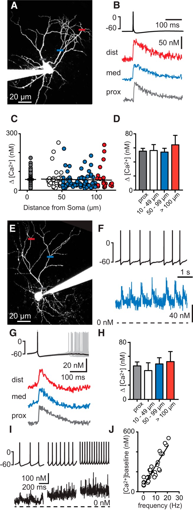
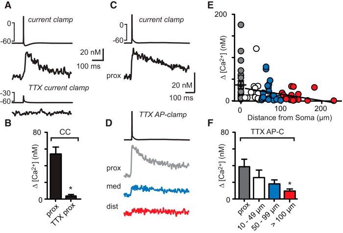
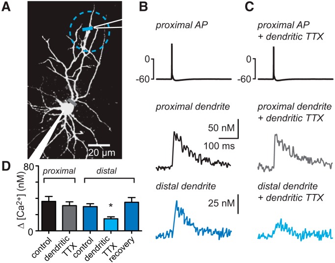
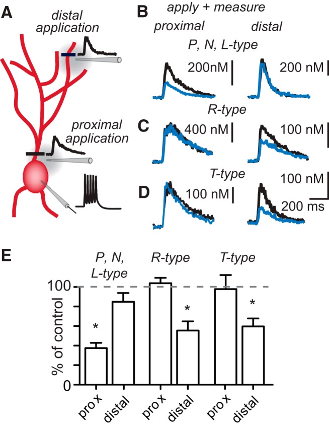
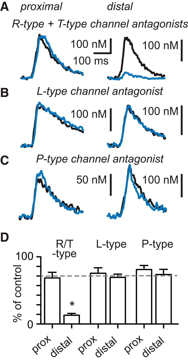
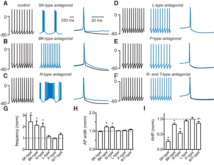
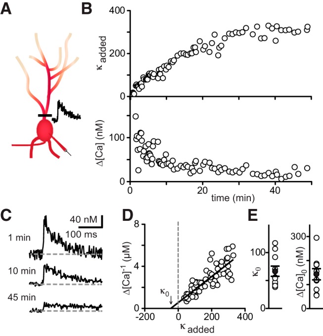
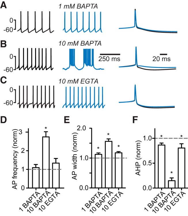
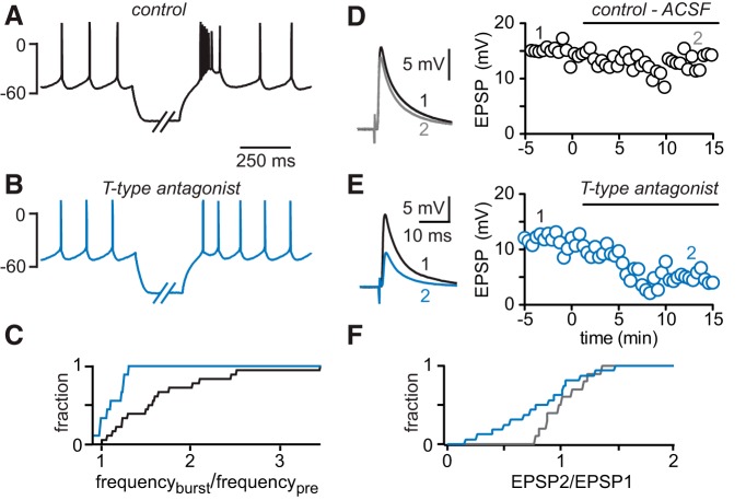
Interneurons are essential to controlling excitability, timing, and synaptic integration in neuronal networks. Golgi cells (GoCs) serve these roles at the input layer of the cerebellar cortex by releasing GABA to inhibit granule cells (grcs). GoCs are excited by mossy fibers (MFs) and grcs and provide feedforward and feedback inhibition to grcs. Here we investigate two important aspects of GoC physiology: the properties of GoC dendrites and the role of calcium signaling in regulating GoC spontaneous activity. Although GoC dendrites are extensive, previous studies concluded they are devoid of voltage-gated ion channels. Hence, the current view holds that somatic voltage signals decay passively within GoC dendrites, and grc synapses onto distal dendrites are not amplified and are therefore ineffective at firing GoCs because of strong passive attenuation. Using whole-cell recording and calcium imaging in rat slices, we find that dendritic voltage-gated sodium channels allow somatic action potentials to activate voltage-gated calcium channels (VGCCs) along the entire dendritic length, with R-type and T-type VGCCs preferentially located distally. We show that R- and T-type VGCCs located in the dendrites can boost distal synaptic inputs and promote burst firing. Active dendrites are thus critical to the regulation of GoC activity, and consequently, to the processing of input to the cerebellar cortex. In contrast, we find that N-type channels are preferentially located near the soma, and control the frequency and pattern of spontaneous firing through their close association with calcium-activated potassium (KCa) channels. Thus, VGCC types are differentially distributed and serve specialized functions within GoCs.

Significance statement: Interneurons are essential to neural processing because they modulate excitability, timing, and synaptic integration within circuits. At the input layer of the cerebellar cortex, a single type of interneuron, the Golgi cell (GoC), carries these functions. The extent of inhibition depends on both spontaneous activity of GoCs and the excitatory synaptic input they receive. In this study, we find that different types of calcium channels are differentially distributed, with dendritic calcium channels being activated by somatic activity, boosting synaptic inputs and enabling bursting, and somatic calcium cannels promoting regular firing. We therefore challenge the current view that GoC dendrites are passive and identify the mechanisms that contribute to GoCs regulating the flow of sensory information in the cerebellar cortex.

Keywords: calcium buffering; calcium channels; calcium-activated potassium channels; cerebellum; dendritic excitability; interneuron.


Conflict of interest statement

The authors declare no competing financial interests.

Figures

Figure 1.
Figure 1.
Action potential-dependent calcium transients in Golgi cell dendrites. A, Two-photon image of a GoC filled with Alexa 594 and the calcium-sensitive dye Fluo-5F. Horizontal bars represent the locations of line scans along the dendrite. B, Top trace, Action potential evoked by a somatic current injection. Bottom traces, Average action potential-dependent calcium transients recorded at the locations indicated in A. C, Amplitudes of dendritic calcium transients dependent on distance from the soma. D, Summary data of 77 cells with calcium transient amplitudes binned according to distance from soma. E, Two-photon image of a spontaneously active GoC with locations of line scans indicated. F, Top trace, Spontaneous action potentials recorded at the soma. Bracket indicates 0 mV and −60 mV. Bottom trace, Calcium signals recorded at a dendritic location indicated in E. Broken line indicates 0 nm baseline calcium. G, Top traces, Aligned spontaneous action potentials (gray traces) and average membrane potential (black trace). Bottom, Average calcium transients in response to spontaneous action potentials recorded at locations indicated in E. H, Summary data of 23 cells. Binned amplitudes of calcium transients. I, Example traces of a GoC firing at different frequencies and associated dendritic calcium signals. Broken line indicates 0 nm baseline calcium. J, Amplitudes of dendritic baseline calcium concentration for different firing frequencies.
Figure 2.
Figure 2.
Dendritic sodium channels promote dendritic calcium signals. A, Top traces, The somatic action potential evokes a calcium transient in the proximal dendrite. Bottom, TTX abolishes both the action potential and the calcium signal. B, Summary data of calcium signals in the presence and in the absence of TTX. C, Waveform of the somatically evoked action potential in current-clamp configuration and the resulting calcium transient in the proximal dendrite. D, Top, Action potential waveform fed back to the GoC in voltage-clamp configuration in the presence of TTX. Bottom, Calcium transients measured at proximal (gray), medial (42 μm, blue), and distal (112 μm, red) dendritic locations. E, The amplitude of calcium transients in dependence on distance from the soma evoked in AP-clamp configuration. F, Binned summary data of calcium transient amplitudes. *Denotes statistical significance.
Figure 3.
Figure 3.
Local inhibition of dendritic sodium channels attenuates dendritic calcium signals. A, Schematic of the experimental setup. Action potentials are evoked somatically, whereas calcium transients are recorded at a proximal and a distal dendritic location. The puff pipette (white lines) is placed near the distal dendrite, and TTX is applied locally (cyan dashed circle). B, Somatically evoked action potential, proximal calcium transient (black), and distal calcium transient (recorded ∼72 μm from soma, blue). C, Somatic action potential during dendritic TTX application, proximal calcium transient (gray), and distal calcium transient (cyan). D, Summary data of calcium signals. *Denotes statistical significance.
Figure 4.
Figure 4.
Different calcium channels mediate proximal and distal calcium transients. A, Schematic of experiment. Bursts of action potentials are evoked somatically. Calcium transients are recorded at a proximal and a distal location; specific calcium channel antagonists are applied locally. B–D, Calcium transients in the presence and absence of calcium channel antagonists applied to the proximal and distal dendrites. B, Antagonists of P-type (ω-agatoxin IVA), N-type (ω-conotoxin GVIA), and L-type (nimodipine) channels were coapplied. C, Calcium transients in the presence and absence of the R-type channel antagonist SNX-482. D, Application of the T-type channel antagonist TTA-P2. E, Summary data of calcium channel pharmacology. *Denotes statistical significance.
Figure 5.
Figure 5.
Bath application of R-type and T-type but not L-type or P-type channel blockers suppresses dendritic calcium signals. A, Coapplication of R-type and T-type channel antagonists does not affect the proximal calcium transient but diminishes the distal dendritic calcium transient. B, Block of L-type calcium channels or (C) P-type calcium channels has no effect on the amplitude of the proximal and distal calcium transients. D, Summary data of the effect of calcium channel antagonists on the amplitude of dendritic calcium signals. *Denotes statistical significance.
Figure 6.
Figure 6.
The effect of calcium-dependent potassium channel and calcium channel inhibition on Golgi cell firing. A, Spontaneously firing GoC in the absence (black trace, left) or in the presence of the SK-type potassium channel blocker apamin (blue trace, right). B, Same experiment but blue trace in the presence of the BK-type potassium channel blocker paxilline. C, The N-type channel blocker ω-conotoxin GVIA. D, The L-type calcium channel antagonist nimodipine. E, The P-type calcium channel blocker ω-agatoxin IVA. F, The R-type and T-type channel blockers SNX-482 and TTA-P2, respectively. G, Summary data of action potential frequency. H, Action potential (AP) width. I, Afterhyperpolarization (AHP). *Denotes statistical significance.
Figure 7.
Figure 7.
Low endogenous calcium buffer capacity in Golgi cells. A, Schematic showing experimental setup for measuring calcium buffer capacity. Action potential-evoked calcium transients are measured continuously at a proximal dendritic site until the calcium-insensitive red dye has equilibrated. B, Top, Added buffer capacity. Bottom, Peak calcium concentration in response to a single action potential during loading of the cell with Alexa 594 and Fluo-5F. C, Calcium transients recorded at time points indicated. D, Relationship between Δ[Ca2+]−1 and added buffer capacity κ during dye loading. Arrow indicates endogenous buffer capacity. E, Summary data of endogenous buffer capacity and amplitude of the calcium signal in the absence of exogenous buffer.
Figure 8.
Figure 8.
The effect of exogenous calcium buffers on firing pattern and action potential waveform in Golgi cells. A–C. Left, Spontaneous action potentials directly after break-in (black traces) and after introduction of the indicated concentrations of exogenous buffer (blue traces). Right, Average action potentials on an expanded time scale. D, Normalized action potential firing rate (left). E, Action potential width. F, Afterhyperpolarization after introduction of exogenous buffers. *Denotes statistical significance.
Figure 9.
Figure 9.
Dendritic T-type calcium channels promote burst firing and boost parallel fiber input to Golgi cells. A, Rebound burst firing following hyperpolarization of a spontaneously active GoC (black trace). B, The T-type channel antagonist TTA-P2 decreases rebound bursting (blue trace). C, Cumulative probability histogram of the ratio of burst and prehyperpolarization action potential frequency (frequburst/frequpre) before (black trace) and after TTA-P2 application (blue trace). D, PF-EPSPs (left, black and gray trace) recorded at the time points indicated (right graphs) in the absence of the T-type channel antagonist. E, PF-EPSPs before (left, black trace) and after TTA-P2 application (blue trace). Inhibition of T-type calcium channels decreases PF-EPSP amplitude in a representative experiment (right). F, Cumulative probability histogram of the EPSP2/EPSP1 ratio: gray trace represents ACSF control; blue trace represents TTA-P2 application.

Similar articles

Cited by

References

    1. Abrahamsson T, Cathala L, Matsui K, Shigemoto R, Digregorio DA. Thin dendrites of cerebellar interneurons confer sublinear synaptic integration and a gradient of short-term plasticity. Neuron. 2012;73:1159–1172. doi: 10.1016/j.neuron.2012.01.027. - DOI - PubMed
    1. Acuna-Goycolea C, Brenowitz SD, Regehr WG. Active dendritic conductances dynamically regulate GABA release from thalamic interneurons. Neuron. 2008;57:420–431. doi: 10.1016/j.neuron.2007.12.022. - DOI - PubMed
    1. Adler EM, Augustine GJ, Duffy SN, Charlton MP. Alien intracellular calcium chelators attenuate neurotransmitter release at the squid giant synapse. J Neurosci. 1991;11:1496–1507. - PMC - PubMed
    1. Aizenman CD, Manis PB, Linden DJ. Polarity of long-term synaptic gain change is related to postsynaptic spike firing at a cerebellar inhibitory synapse. Neuron. 1998;21:827–835. doi: 10.1016/S0896-6273(00)80598-X. - DOI - PubMed
    1. Alle H, Kubota H, Geiger JR. Sparse but highly efficient Kv3 outpace BKCa channels in action potential repolarization at hippocampal mossy fiber boutons. J Neurosci. 2011;31:8001–8012. doi: 10.1523/JNEUROSCI.0972-11.2011. - DOI - PMC - PubMed

Publication types

Substances

LinkOut - more resources