

Nanoparticle Probes for Structural and Functional Photoacoustic Molecular Tomography
- PMID: 26609534
- PMCID: PMC4644549
- DOI: 10.1155/2015/757101
Nanoparticle Probes for Structural and Functional Photoacoustic Molecular Tomography
Abstract
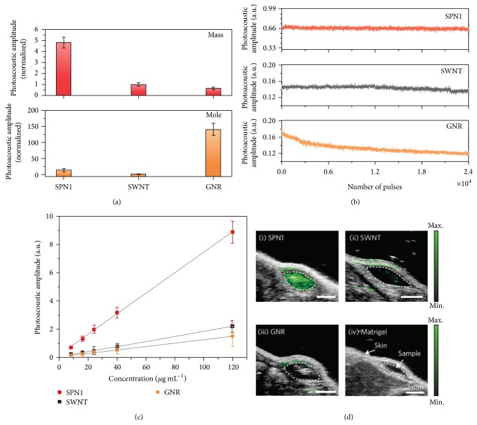
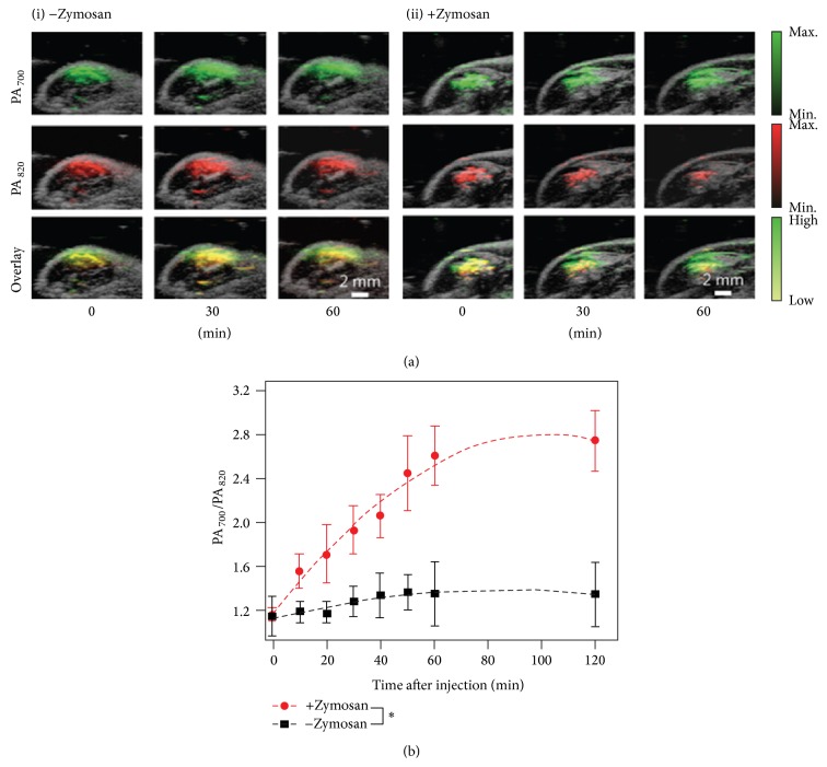
Nowadays, nanoparticle probes have received extensive attention largely due to its potential biomedical applications in structural, functional, and molecular imaging. In addition, photoacoustic tomography (PAT), a method based on the photoacoustic effect, is widely recognized as a robust modality to evaluate the structure and function of biological tissues with high optical contrast and high acoustic resolution. The combination of PAT with nanoparticle probes holds promises for detecting and imaging diseased tissues or monitoring their treatments with high sensitivity. This review will introduce the recent advances in the emerging field of nanoparticle probes and their preclinical applications in PAT, as well as relevant perspectives on future development.
Figures
Similar articles
-
Recent progress in photoacoustic molecular imaging.Curr Opin Chem Biol. 2018 Aug;45:104-112. doi: 10.1016/j.cbpa.2018.03.016. Epub 2018 Apr 7. Curr Opin Chem Biol. 2018. PMID: 29631120 Free PMC article. Review.
-
Photoacoustic tomography: principles and advances.Electromagn Waves (Camb). 2014;147:1-22. doi: 10.2528/pier14032303. Electromagn Waves (Camb). 2014. PMID: 25642127 Free PMC article.
-
Structural and functional photoacoustic molecular tomography aided by emerging contrast agents.Chem Soc Rev. 2014;43(20):7132-70. doi: 10.1039/c4cs00086b. Chem Soc Rev. 2014. PMID: 24967718 Free PMC article. Review.
-
Photoacoustic Probes for Molecular Detection: Recent Advances and Perspectives.Small. 2018 Jul;14(30):e1800782. doi: 10.1002/smll.201800782. Epub 2018 Jun 5. Small. 2018. PMID: 29873182 Review.
-
Photoacoustic sentinel lymph node imaging with self-assembled copper neodecanoate nanoparticles.ACS Nano. 2012 Feb 28;6(2):1260-7. doi: 10.1021/nn203895n. Epub 2012 Jan 20. ACS Nano. 2012. PMID: 22229462 Free PMC article.
Cited by
-
Big Potential from Small Agents: Nanoparticles for Imaging-Based Companion Diagnostics.ACS Nano. 2018 Mar 27;12(3):2106-2121. doi: 10.1021/acsnano.7b07252. Epub 2018 Mar 1. ACS Nano. 2018. PMID: 29462554 Free PMC article. Review.
-
Towards optimized naphthalocyanines as sonochromes for photoacoustic imaging in vivo.Photoacoustics. 2018 Jan 11;9:49-61. doi: 10.1016/j.pacs.2017.12.001. eCollection 2018 Mar. Photoacoustics. 2018. PMID: 29707479 Free PMC article.
-
Advanced optoacoustic methods for multiscale imaging of in vivo dynamics.Chem Soc Rev. 2017 Apr 18;46(8):2158-2198. doi: 10.1039/c6cs00765a. Chem Soc Rev. 2017. PMID: 28276544 Free PMC article. Review.
-
Iron oxide nanoparticles as multimodal imaging tools.RSC Adv. 2019 Dec 6;9(69):40577-40587. doi: 10.1039/c9ra08612a. eCollection 2019 Dec 3. RSC Adv. 2019. PMID: 35542631 Free PMC article. Review.
-
Semiconducting Polymer Dots for Point-of-Care Biosensing and In Vivo Bioimaging: A Concise Review.Biosensors (Basel). 2023 Jan 14;13(1):137. doi: 10.3390/bios13010137. Biosensors (Basel). 2023. PMID: 36671972 Free PMC article. Review.
References
-
- Wang Y., Xie X., Wang X., et al. Photoacoustic tomography of a nanoshell contrast agent in the in vivo rat brain. Nano Letters. 2004;4(9):1689–1692. doi: 10.1021/nl049126a. - DOI
Publication types
MeSH terms
LinkOut - more resources
Full Text Sources
Other Literature Sources
