. 2016 Apr;26(4):360-76.
doi: 10.1093/glycob/cwv108. Epub 2015 Nov 26.

Mucin-type O-glycosylation is controlled by short- and long-range glycopeptide substrate recognition that varies among members of the polypeptide GalNAc transferase family

Affiliations

Mucin-type O-glycosylation is controlled by short- and long-range glycopeptide substrate recognition that varies among members of the polypeptide GalNAc transferase family

Leslie Revoredo et al. Glycobiology. 2016 Apr.

Abstract

A large family of UDP-GalNAc:polypeptide GalNAc transferases (ppGalNAc-Ts) initiates and defines sites of mucin-type Ser/Thr-O-GalNAc glycosylation. Family members have been classified into peptide- and glycopeptide-preferring subfamilies, although both families possess variable activities against glycopeptide substrates. All but one isoform contains a C-terminal carbohydrate-binding lectin domain whose roles in modulating glycopeptide specificity is just being understood. We have previously shown for several peptide-preferring isoforms that the presence of a remote Thr-O-GalNAc, 6-17 residues from a Ser/Thr acceptor site, may enhance overall catalytic activity in an N- or C-terminal direction. This enhancement varies with isoform and is attributed to Thr-O-GalNAc interactions at the lectin domain. We now report on the glycopeptide substrate utilization of a series of glycopeptide (human-ppGalNAc-T4, T7, T10, T12 and fly PGANT7) and peptide-preferring transferases (T2, T3 and T5) by exploiting a series of random glycopeptide substrates designed to probe the functions of their catalytic and lectin domains. Glycosylation was observed at the -3, -1 and +1 residues relative to a neighboring Thr-O-GalNAc, depending on isoform, which we attribute to specific Thr-O-GalNAc binding at the catalytic domain. Additionally, these glycopeptide-preferring isoforms show remote lectin domain-assisted Thr-O-GalNAc enhancements that vary from modest to none. We conclude that the glycopeptide specificity of the glycopeptide-preferring isoforms predominantly resides in their catalytic domain but may be further modulated by remote lectin domain interactions. These studies further demonstrate that both domains of the ppGalNAc-Ts have specialized and unique functions that work in concert to control and order mucin-type O-glycosylation.

Keywords: glycoprotein biosynthesis; glycosyltransferase; lectin; mucin.


Figures

Fig. 1.
Fig. 1.
Phylogenetic organization and structure of the ppGalNAc-T's. (A) Phylogenetic tree of the ppGalNAc-T (h-GALNT) gene family (modified from Bennett et al. 2012). “Peptide-preferring” transferases in subfamilies Ia–Ig (except Ie) are highlighted in red. The “glycopeptide-preferring” transferases (subfamilies IIa–IIb) are highlighted in blue. Note that subfamily Ie (“Y” series of transferases (Li et al. 2012)) is highlighted in gray due to their largely unknown activities. (B) ppGalNAc-T2 crystal structure (PDP 2FFU) with EA2 peptide bound to the catalytic domain (Fritz et al. 2006). The residues of EA2 (5STTPAPTTK13) are space-filled: the N-terminal Ser is blue; the C-terminal Lys is red; the Pro residues are purple and the remaining Thr are brown. In the lectin domain, the Asp458 residue that is required for glycopeptide binding is space-filled in pink (Wandall et al. 2007; Pederson et al. 2011). Figure (B) from Gerken et al. (2013).
Fig. 2.
Fig. 2.
Activities of the ppGalNAc-T's against glycopeptide substrates showing GP(T*10)C as the preferred substrate for the “glycopeptide-preferring” ppGalNAc-Ts. Representative time course plots of net UDP-[3H]-GalNAc utilization (i.e. GalNAc transfer to peptide and water) for the “peptide-preferring” ppGalNAc-T5 (A) and “glycopeptide-preferring” ppGalNAc-T4 (B), T12 (C), T10 (D), T7 (E) and PGANT7 (F). Shown are plots for the random glycopeptide substrates GP(T*22)R (red diamond), GP(A22)R (pink upward triangle), GP(T*10)L (blue square), GP(A10)L (turquoise downward triangle) and GP(T*10)C (green circle) glycosylated by the indicated transferase. These plots represent the mole fraction of [3H]-GalNAc (50 µM) transferred to both glycopeptide substrate (1.5–1.7 mM) and water as a function of time as described in Experimental Procedures. (Standard deviation values for all time points were typically less than ∼0.05, although SD values of ∼0.15 were obtained for GP(T*10)C against the overnight time points for ppGalNAc-T5 and T12). These plots represent the average values from 2 to 4 experimental runs.
Fig. 3.
Fig. 3.
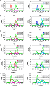
Sephadex G10 gel filtration chromatograms demonstrating elevated GalNAc hydrolysis (transfer to water) for the “glycopeptide-preferring” ppGalNAc-Ts. Chromatograms are shown for the “peptide-preferring” ppGalNAc-T5 (A) and “glycopeptide-preferring” ppGalNAc-T4 (B), T12 (C), T10 (D), T7 (E) and the fly PGANT7 (F). Left panels are chromatograms for random (glyco)peptide substrates GP(T*22)R (red diamond), GP(A22)R (pink upward triangle) and GP(T*10)C (green circle) while the right panels show chromatograms for GP(T*10)L (blue square), GP(A10)L (turquoise downward triangle) and GP(T*10)C (green circle). These plots demonstrate the incorporation of [3H]-GalNAc into (glyco)peptide substrate (fractions 24–31) and hydrolysis (i.e. transfer to water, fractions 36–43) for the overnight incubations (1200 min) shown in Figure 2. These plots are representative plots from a single experiment, although two or more experiments displaying nearly identical results were performed.
Fig. 4.
Fig. 4.
Efficiency of GalNAc transfer to acceptor glycopeptides varies with ppGalNAc-T family. Bars represent the percent (i.e. efficiency) of GalNAc transfer to the indicated glycopeptide substrates: GP(T*22)R, GP(T*10)L and GP(T*10)C respectively. Left grouping of bars represents the “peptide-preferring” subfamily I transferases, ppGalNAc-T1, T2, T3, T5, T13 and T16, while the right grouping of bars represents the “glycopeptide-preferring” subfamily II transferases ppGalNAc-T4, T12, T10, T7 and PGANT7. Values are obtained from 2 to 4 G10 gel filtration runs of standard overnight incubations (in Figure 3 and data not shown) and (Gerken et al. 2013). Note that these values are displayed to reveal trends and may vary with the transferase reaction conditions. Error bars represent SD from 2 to 4 experiments. This figure is available in black and white in print and in color at Glycobiology online.
Fig. 5.
Fig. 5.
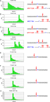
[3H]-GalNAc incorporation into the X acceptor residues of GP(T*10)C revealing specific sites of glycosylation that vary with isoform. Edman amino acid sequence analysis of [3H]-GalNAc incorporation into GP(T*10)C by ppGalNAc-T2 (A), T3 (B), T5 (C), T4 (D), T12 (E), T10 (F), T7 (G) and the fly PGANT7 (H). At the right of each panel is representations depicting major sites of glycosylation (arrows) for GP(T*10)C. Below these representations, for panels A–D, representations depicting the sites of glycosylation (X's and arrows) of the lectin probing glycopeptides GP(T*22)R and GP(T*10)L are given (determined previously (Gerken et al. 2013) or from this work). All plots are for overnight incubations of GP(T*10)C with the indicated transferase and are representative of at least two sequence determinations. This figure is available in black and white in print and in color at Glycobiology online.
Fig. 6.
Fig. 6.
Glycosylation of the GP(T*10)C control substrate, GP(A10)C, demonstrating the role of T* in site selection. Sephadex G10 gel filtration analysis (left panels) and Edman amino acid sequence analysis of [3H]-GalNAc incorporation into GP(A10)C (right panels) for ppGalNAc-T2 (A), T4 (B) and T10 (C). Plots in the left panels are for overnight incubations of GP(A10)C and GP(T*10)C while the right panel plots are for overnight incubation of GP(A10)C. Note the broad peak of [3H] incorporation with ppGalNAc-T2 (right panel) and the loss of [3H] incorporation with ppGalNAc-T4 and T10 (left and right panels) into GP(A10)C compared with GP(T*10)C given in Figure 5A, D and F. These plots are determined from a single experimental run for each transferase. This figure is available in black and white in print and in color at Glycobiology online.
Fig. 7.
Fig. 7.
[3H]-GalNAc incorporation into the X acceptor residues of glycopeptide substrates GP(T*22)R and GP(T*10)L for the “glycopeptide-preferring” ppGalNAc-Ts showing isoform-specific long-range N- and/or C-terminal specificity. Edman amino acid sequence analysis of the incorporation of [3H]-GalNAc into GP(T*22)R (left panels) and GP(T*10)L (right panels) for overnight incubations with ppGalNAc-T4 (A), T12 (B), T7 (C) and the fly PGANT7 (D). Plots are representative of 1–2 sequence determinations. This figure is available in black and white in print and in color at Glycobiology online.
Fig. 8.
Fig. 8.
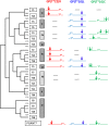
Correlation of glycopeptide specificities with the ppGalNAc-T phylogenetic family tree. To the right of the ppGalNAc-T subfamily phylogenetic tree (Bennett et al. 2012) are plotted the glycopeptide preferences obtained from this work and previous work (Gerken et al. 2013). Glycopeptide substrate cartoons representing GP(T*22)R (left column), GP(T*10)L (middle column) and GP(T*10)C (right column) are shown where the T* represents the position of the initial Ser/Thr-O-GalNAc while the arrows indicate the position(s) glycosylated by the indicated transferase isoform. Note for GP(T*22)R and GP(T*10)L the arrows represent a broad distribution of glycosylation. Substrates with low or no activities are marked with a thin black line. Transferases whose preferences have not been determined against these substrates are left as a blank space. GP(T*10)C cartoons having arrows with parenthesis represent the proposed +/−5 lectin domain-mediated glycosylation site preference. This figure is available in black and white in print and in color at Glycobiology online.
Fig. 9.
Fig. 9.
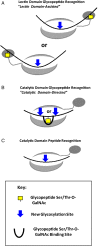
Schematic representations of catalytic domain-directed and lectin domain-assisted glycopeptide activities of the ppGalNAc-Ts. (A) Lectin domain glycopeptide recognition (i.e. the lectin domain-assisted property) mediated by the binding of T* or S* at the lectin domain (upper panel ppGalNAc-T1, T2, T5, T13, T14, T16 and PGANT7, lower panel ppGalNAcT3, T4, T5, T6, T12, T13, T16 and PAGANT7). (B) Catalytic domain glycopeptide recognition (i.e. the catalytic-domain directed property) by the mixed (glyco)peptide-preferring subfamily IIa transferases (ppGalNAc-T4 and T12) and the strict glycopeptide-preferring subfamily IIb (ppGalNAc-T7, T10 and fly PGANT7). (C) Peptide substrate binding to the catalytic domain directing glycosylation by the remote glycopeptide/peptide-preferring subfamilies Ia–Id (ppGalNAc-T1, T2, T3, T5, T6, T13, T14 and T16) and mixed (glyco)peptide-preferring subfamily IIa (ppGalNAc-T4 and T12). The large gray oval represents the catalytic domain while the smaller right or left tethered ovals represent the lectin domain and the thick black line represents (glyco)peptide acceptor (see Discussion for an explanation of the renamed family/subfamily classifications). This figure is available in black and white in print and in color at Glycobiology online.

References

    1. Barb AW, Meng L, Gao Z, Johnson RW, Moremen KW, Prestegard JH. 2012. NMR characterization of immunoglobulin G Fc glycan motion on enzymatic siaylation. Biochemistry. 51:4618–4626. - PMC - PubMed
    1. Bennett EP, Hassan H, Hollingsworth MA, Clausen H. 1999. A novel human UDP-N-acetyl-D-galactosamine:polypeptide N-acetylgalactosaminyltransferase, GalNAc-T7, with specificity for partial GalNAc-glycosylated acceptor substrates. FEBS Lett. 460:226–230. - PubMed
    1. Bennett EP, Hassan H, Mandel U, Mirgorodskaya E, Roepstorff P, Burchell J, Taylor-Papadimitriou J, Hollingsworth MA, Merkx G, van Kessel AG et al. . 1998. Cloning of a human UDP-N-acetyl-alpha-D-galactosamine:polypeptide N-acetylgalactosaminyltransferase that complements other GalNAc-transferases in complete O-glycosylation of the MUC1 tandem repeat. J Biol Chem. 273:30472–30481. - PubMed
    1. Bennett EP, Mandel U, Clausen H, Gerken TA, Fritz TA, Tabak LA. 2012. Control of mucin-type O-glycosylation: A classification of the polypeptide GalNAc-transferase gene family. Glycobiology. 22:736–756. - PMC - PubMed
    1. Berois N, Mazal D, Ubillos L, Trajtenberg F, Nicolas A, Sastre-Garau X, Magdelenat H, Osinaga E. 2006. UDP-N-acetyl-D-galactosamine: Polypeptide N-acetylgalactosaminyltransferase-6 as a new immunohistochemical breast cancer marker. J Histochem Cytochem. 54:317–328. - PubMed

Publication types

MeSH terms