Review
. 2015:76:305-36.
doi: 10.1016/bs.ctm.2015.07.001. Epub 2015 Jul 30.

Building from the Ground up: Basement Membranes in Drosophila Development

Affiliations
Review

Building from the Ground up: Basement Membranes in Drosophila Development

Adam J Isabella et al. Curr Top Membr. 2015.

Abstract

Basement membranes (BMs) are sheetlike extracellular matrices found at the basal surfaces of epithelial tissues. The structural and functional diversity of these matrices within the body endows them with the ability to affect multiple aspects of cell behavior and communication; for this reason, BMs are integral to many developmental processes. The power of Drosophila genetics, as applied to the BM, has yielded substantial insight into how these matrices influence development. Here, we explore three facets of BM biology to which Drosophila research has made particularly important contributions. First, we discuss how newly synthesized BM proteins are secreted to and assembled exclusively on basal epithelial surfaces. Next, we examine how regulation of the structural properties of the BM mechanically supports and guides tissue morphogenesis. Finally, we explore how BMs influence development through the modulation of several major signaling pathways.

Keywords: Basement membrane; Development; Drosophila; Morphogenesis; Secretion; Signaling.


Figures

Figure 1
Figure 1
Overview of the core BM proteins in Drosophila
Figure 2
Figure 2. Cellular sources of BM proteins in Drosophila
BM proteins are synthesized and secreted by three primary cell types. 1: Synthesis and local deposition by hemocytes. 2: Synthesis by and long range diffusion from the fat body. 3: Synthesis and secretion by the epithelium itself.
Figure 3
Figure 3. Local synthesis and polarized secretion of Collagen IV in the follicular epithelium
Within the follicular epithelium, Collagen IV transcripts accumulate basally and are transcribed into a basal region of the ER. ER resident proteins assist in the folding and packaging of Collagen IV for transport to the Golgi. After Collagen IV transits through the Golgi, Crag and Rab10 promote delivery of Collagen IV-containing exocytic vesicles to basal regions of the plasma membrane and prevent Rab11-dependent targeting of Collagen IV-containing vesicles to the apical surface. Inset: blow-up of indicated region.
Figure 4
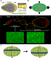
Figure 4. BM function in egg chamber elongation
(A) Illustration of a transverse section through an egg chamber. Central germ cells are surrounded by a somatic epithelium of follicle cells, which assemble a BM on their basal surfaces. (B) Model of the molecular corset. Polarized linear fibril-like structures in the BM are hypothesized to constrain egg chamber growth in the direction of polarization, biasing growth to occur along the A-P axis. Arrows indicate direction and magnitude of growth. (C) BM structural dynamics during elongation. Top: a developmental array of egg chambers showing cell outlines (actin) and the BM (Viking-GFP). Bottom: fluorescent micrographs of Collagen IV (Viking-GFP) in the BM. Young, round egg chambers exhibit no obvious BM structure, while older, elongating egg chambers display polarized fibrils within the BM. (D) Overview of egg chamber rotation. In this illustration, the BM is partially transparent to reveal the cells underneath. The dark row of cells in each image represents the same cells at two different time points. The egg chamber rotates within a stationary BM, in the direction of BM fibrils. Curved arrows indicate direction of egg chamber rotation.
Figure 5
Figure 5. BM regulation of Dpp signaling in three developmental contexts
(A) Embryonic Malpighian tubule morphogenesis. Diffusible Dpp protein attracts the growing Malpighian tubule. Hemocyte-deposited Collagen IV around the Malpighian tubule promotes reception of the Dpp signal by tubule cells. (B) Ovarian germline stem cell (GSC) maintenance. GSCs are maintained within the stem cell niche via interaction with cap cells. Collagen IV in the BM and the HSPG Dally concentrate cap cell-derived Dpp to promote signal reception by GSCs but not differentiating daughter cells. (C) Intestinal stem cell (ISC) maintenance. ISCs exhibit basally positioned cell bodies that adhere to the BM. Dpp is concentrated by Collagen IV within the BM to promote a high level of signal reception by ISCs but not the more apically-localized, differentiating enteroblasts.

References

    1. Andrew DJ, Ewald AJ. Morphogenesis of epithelial tubes: Insights into tube formation, elongation, and elaboration. Developmental Biology. 2010;341(1):34–55. - PMC - PubMed
    1. Anitei M, Hoflack B. Exit from the trans-Golgi network: from molecules to mechanisms. Current Opinion in Cell Biology. 2011;23(4):443–51. - PubMed
    1. Babbey C, Ahktar N, Wang E, Chen C, Grant B, Dunn K. Rab10 Regulates Membrane Transport through Early Endosomes of Polarized Madin-Darby Canine. Molecular Biology of. 2006;17:3156–3175. - PMC - PubMed
    1. Balla T. Phosphoinositides: tiny lipids with giant impact on cell regulation. Physiological Reviews. 2013;93(3):1019–137. - PMC - PubMed
    1. Barr FA. Rab GTPases and membrane identity: Causal or inconsequential? The Journal of Cell Biology. 2013;202(2):191–9. - PMC - PubMed

Publication types

LinkOut - more resources