. 2016 Feb;7(1):77-86.
doi: 10.1007/s13244-015-0446-5. Epub 2015 Nov 26.

Ultrasonography of thyroid nodules: a pictorial review

Affiliations

Ultrasonography of thyroid nodules: a pictorial review

Cheng Xie et al. Insights Imaging. 2016 Feb.

Abstract

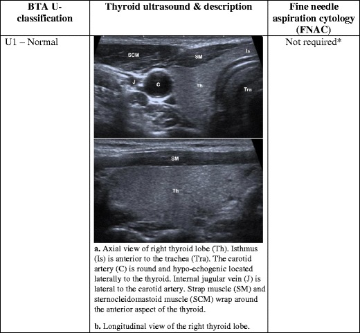
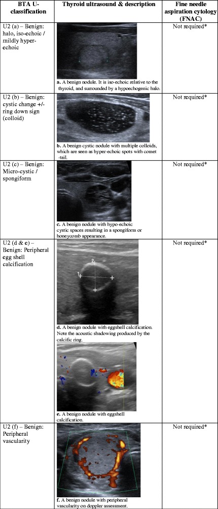
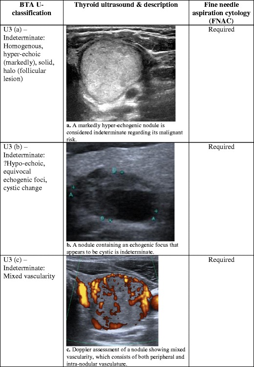
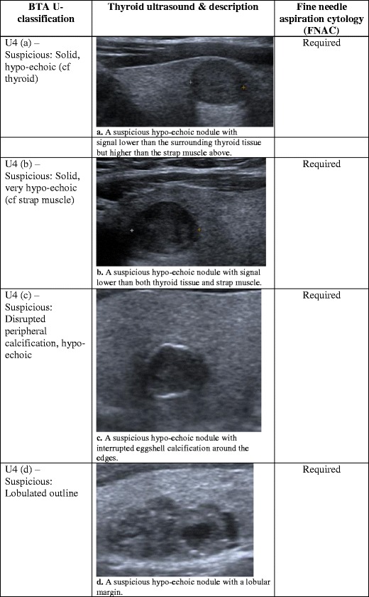
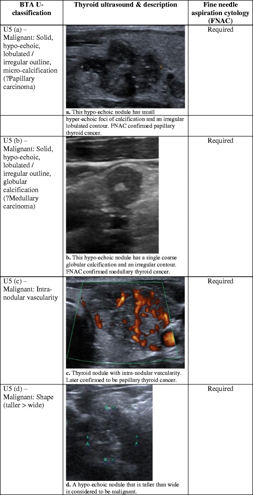
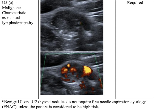
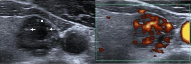
Thyroid nodules are a common occurrence in the general population, and these incidental thyroid nodules are often referred for ultrasound (US) evaluation. US provides a safe and fast method of examination. It is sensitive for the detection of thyroid nodules, and suspicious features can be used to guide further investigation/management decisions. However, given the financial burden on the health service and unnecessary anxiety for patients, it is unrealistic to biopsy every thyroid nodule to confirm diagnosis. The British Thyroid Association (BTA) has recently produced a US classification (U1-U5) of thyroid nodules to facilitate the decision-making process regarding the need to perform fine-needle aspiration cytology (FNAC) for suspicious cases. In this pictorial review, we provide a complete series of sonographic images to illustrate benign and malignant features of thyroid nodules according to the U1-5 classification. Specifically, we highlight morphologic characteristic of the nodule, including its echo signal in relation to its consistency, nodular size, number and contour. Additional diagnostic features such as halo, colloid, calcification and vascular patterns are also discussed in detail. The aim is to assist radiologists and clinicians in recognising sonographic patterns of benign, suspicious and malignant nodules based on U1-5 criteria, and in planning for further investigations.

Main messages: • Ultrasound is sensitive in identifying suspicious features, which require aspiration. • Whether nodules require aspiration should be based on sonographic features and clinical findings. • U1-5 classification of sonographic findings can help determine whether aspiration is necessary.

Keywords: Fine-needle aspiration; Sonographic features; Thyroid cancer; Thyroid nodule; Ultrasound.


Figures

Fig. 1
Fig. 1
(a) Axial view of right thyroid lobe (Th). Isthmus (Is) is anterior to the trachea (Tra). The carotid artery (C) is round and hypo-echogenic located laterally to the thyroid. The internal jugular vein (J) is lateral to the carotid artery. The strap muscle (SM) and sternocleidomastoid muscle (SCM) wrap around the anterior aspect of the thyroid. (b) Longitudinal view of the right thyroid lobe
Fig. 2
Fig. 2
(a) A benign nodule. It is iso-echoic relative to the thyroid, and surrounded by a hypo-echogenic halo. (b) A benign cystic nodule with multiple colloids, which are seen as hyper-echoic spots with comet-tail. (c) A benign nodule with hypo-echoic cystic spaces resulting in a spongiform or honeycomb appearance. (d) A benign nodule with eggshell calcification. Note the acoustic shadowing produced by the calcific ring. (e) A benign nodule with eggshell calcification. (f) A benign nodule with peripheral vascularity on Doppler assessment
Fig. 3
Fig. 3
(a) A markedly hyper-echogenic nodule is considered indeterminate regarding its risk of malignancy. (b) A nodule containing an echogenic focus that appears to be cystic is indeterminate. (c) Doppler assessment of a nodule showing mixed vascularity, which consists of both peripheral and intra-nodular vasculature
Fig. 4
Fig. 4
(a) A suspicious hypo-echoic nodule with signal lower than the surrounding thyroid tissue but higher than the strap muscle above. (b) A suspicious hypo-echoic nodule with signal lower than both thyroid tissue and strap muscle. (c) A suspicious hypo-echoic nodule with interrupted eggshell calcification around the edges. (d) A suspicious hypo-echoic nodule with a lobular margin
Fig. 5
Fig. 5
(a) This hypo-echoic nodule has small hyper-echoic foci of calcification and an irregular lobulated contour. FNAC confirmed papillary thyroid cancer. (b) This hypo-echoic nodule has a single coarse globular calcification and an irregular contour. FNAC confirmed medullary thyroid cancer. (c) Thyroid nodule with intra-nodular vascularity, later confirmed to be papillary thyroid cancer. (d) A hypo-echoic nodule that is taller than wide is considered to be malignant. (e) An abnormal lymph node with malignant features—irregular contours, mixed echotexture and vascularity. An abnormal lymph node would result in a U5 category despite any benign features of the thyroid nodule
Fig. 5
Fig. 5
(a) This hypo-echoic nodule has small hyper-echoic foci of calcification and an irregular lobulated contour. FNAC confirmed papillary thyroid cancer. (b) This hypo-echoic nodule has a single coarse globular calcification and an irregular contour. FNAC confirmed medullary thyroid cancer. (c) Thyroid nodule with intra-nodular vascularity, later confirmed to be papillary thyroid cancer. (d) A hypo-echoic nodule that is taller than wide is considered to be malignant. (e) An abnormal lymph node with malignant features—irregular contours, mixed echotexture and vascularity. An abnormal lymph node would result in a U5 category despite any benign features of the thyroid nodule
Fig. 6
Fig. 6
The thyroid nodule in the left lobe consists of colloids with their characteristic ‘comet-tail’ shadowing (arrows). However, it is hypo-echoic with an echo signal close to the adjacent strap muscle. Power Doppler shows marked intra-nodular vascularity. Multiple sinister characteristics of the nodule put it between U4 and U5, which require FNAC

References

    1. Frates MC, Benson CB, Charboneau JW, Cibas ES, Clark OH, Coleman BG, et al. Management of thyroid nodules detected at US: society of radiologists in ultrasound consensus conference statement. Radiology. 2005;237(3):794–800. doi: 10.1148/radiol.2373050220. - DOI - PubMed
    1. Jemal A, Murray T, Ward E, Samuels A, Tiwari RC, Ghafoor A, et al. Cancer statistics, 2005. CA Cancer J Clin. 2005;55(1):10–30. doi: 10.3322/canjclin.55.1.10. - DOI - PubMed
    1. Hambly NM, Gonen M, Gerst SR, Li D, Jia X, Mironov S, et al. Implementation of evidence-based guidelines for thyroid nodule biopsy: a model for establishment of practice standards. AJR Am J Roentgenol. 2011;196(3):655–60. doi: 10.2214/AJR.10.4577. - DOI - PubMed
    1. Kim EK, Park CS, Chung WY, Oh KK, Kim DI, Lee JT, et al. New sonographic criteria for recommending fine-needle aspiration biopsy of nonpalpable solid nodules of the thyroid. AJR Am J Roentgenol. 2002;178(3):687–91. doi: 10.2214/ajr.178.3.1780687. - DOI - PubMed
    1. Frates MC, Benson CB, Doubilet PM, Kunreuther E, Contreras M, Cibas ES, et al. Prevalence and distribution of carcinoma in patients with solitary and multiple thyroid nodules on sonography. J Clin Endocrinol Metab. 2006;91(9):3411–7. doi: 10.1210/jc.2006-0690. - DOI - PubMed

LinkOut - more resources