Review
. 2016 Feb;65(1):9-22.
doi: 10.1093/jmicro/dfv366. Epub 2015 Nov 25.

Single-particle cryo-electron microscopy of macromolecular complexes

Affiliations
Review

Single-particle cryo-electron microscopy of macromolecular complexes

Georgios Skiniotis et al. Microscopy (Oxf). 2016 Feb.

Abstract

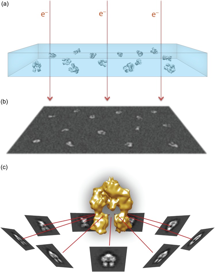
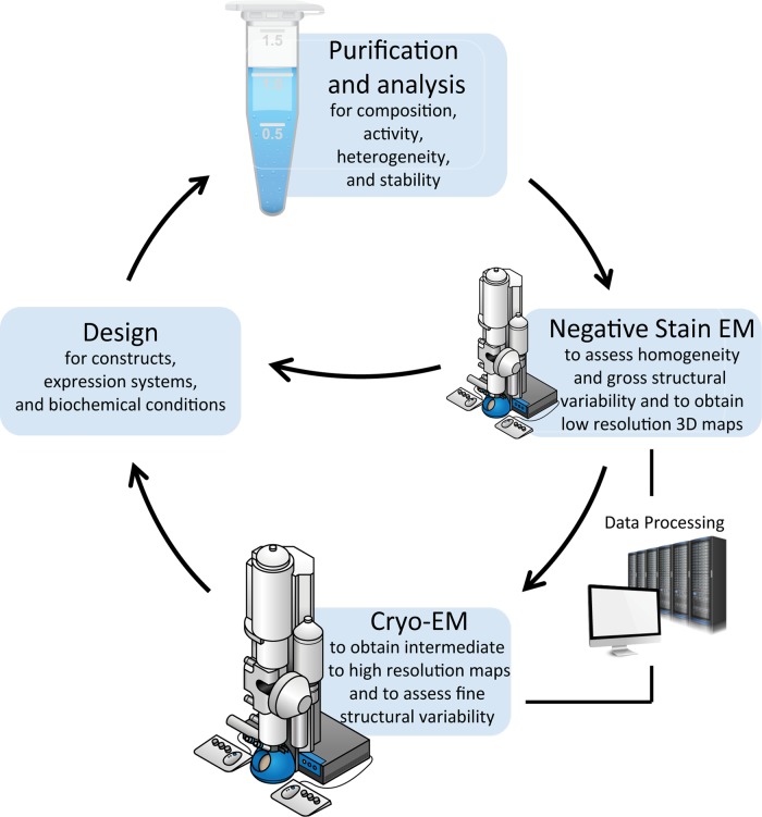
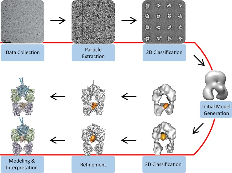
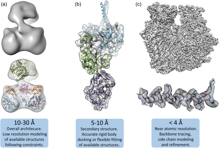
Recent technological breakthroughs in image acquisition have enabled single-particle cryo-electron microscopy (cryo-EM) to achieve near-atomic resolution structural information for biological complexes. The improvements in image quality coupled with powerful computational methods for sorting distinct particle populations now also allow the determination of compositional and conformational ensembles, thereby providing key insights into macromolecular function. However, the inherent instability and dynamic nature of biological assemblies remain a tremendous challenge that often requires tailored approaches for successful implementation of the methodology. Here, we briefly describe the fundamentals of single-particle cryo-EM with an emphasis on covering the breadth of techniques and approaches, including low- and high-resolution methods, aiming to illustrate specific steps that are crucial for obtaining structural information by this method.

Keywords: cryo-EM; macromolecular structure; negative-stain EM; single-particle EM.


Figures

Fig. 1.
Fig. 1.
Overview of single-particle cryo-EM visualization. (a) Purified macromolecular complexes in solution are rapidly frozen in a thin layer of vitreous ice, ideally in random orientations. (b) Electrons passing through the sample in a transmission electron microscope are recorded as 2D projection images on a recording medium, such as a CCD or a direct electron detector. The particle projections have low signal-to-noise ratio due to the relatively weak electron scattering and the low-dose imaging requirements. (c) Similar particle projections are combined to form class averages that can be assigned with experimentally determined angular relationship. The projection averages are back-projected along their assigned angles to generate a 3D density map representing the particles.
Fig. 2.
Fig. 2.
Overview of sample optimization and analysis for cryo-EM. For many samples iterative processes are necessary for obtaining a structure, improving resolution, identifying conformations or validating structural models. Following purification, initial negative-stain EM screening can provide a check-point regarding the suitability of the specimen for cryo-EM studies. Evaluation of samples as heterogeneous or unstable should lead to the redesign of constructs, buffer conditions and purification strategies to facilitate improvement. Significant computational analysis is required throughout EM steps for 2D/3D classification and map refinement. Following an initial cryo-EM characterization, additional construct designs or biochemical conditions are often necessary to probe conformational changes under different biochemical sates (e.g. inactive or activated enzymes or receptors) or validate domain localization in maps at intermediate to low-resolution.
Fig. 3.
Fig. 3.
Overview of steps for the determination of single-particle cryo-EM 3D reconstructions. Cryo-EM images are acquired and individual particles are extracted manually or with automation, ideally followed by user examination to exclude falsely picked projections. The particle projections are subjected to 2D classification and averaging, aiming to group the projections according to their similarity. An initial 3D model may be generated ab initio using class averages representing different particle orientations, by geometrically constrained 3D reconstruction methods such as by RCT, or by utilizing available structural information. The initial 3D model can serve as a reference for 3D classification of the data, a useful step for identifying distinct conformations or macromolecular compositions. The 3D classification also facilitates identification of particle subsets that are amenable to higher resolution refinement. Depending on the achieved resolution, the final maps can be used for de novo structure building or the modeling of available structures based on various criteria.
Fig. 4.
Fig. 4.
Molecular modeling at different resolution ranges. (a) Low resolution maps (10–30 Å), e.g. from a nitric oxide synthase (NOS) complex [69], can be used to dock large domains or protomer subunits and often require significant additional biochemical or structural information to provide constraints and validate fitting. (b) Intermediate resolution 3D reconstructions (5–10 Å), such as the one shown for a polyketide synthase (PKS) module [54], identify secondary structure elements, like α-helices, and subdomain densities that are reliably docked within the density map. Structure docking in this resolution range may benefit from flexible fitting procedures to identify novel conformational arrangements. (c) High-resolution maps (<4 Å), such as from a 70S ribosome (unpublished data), resolve backbone and side-chain densities, enabling de novo structure building.

References

    1. Armache J P, Jarasch A, Anger A M, Villa E, Becker T, Bhushan S, Jossinet F, Habeck M, Dindar G, Franckenberg S, Marquez V, Mielke T, Thomm M, Berninghausen O, Beatrix B, Söding J, Westhof E, Wilson D N, Beckmann R (2010) Cryo-EM structure and rRNA model of a translating eukaryotic 80S ribosome at 5.5-Å resolution. Proc. Natl Acad. Sci. USA 107: 19748–19753. - PMC - PubMed
    1. Ludtke S J, Baker M L, Chen D H, Song J L, Chuang D T, Chiu W (2008) De novo backbone trace of GroEL from single particle electron cryomicroscopy. Structure 16: 441–448. - PubMed
    1. Rabl J, Smith D M, Yu Y, Chang S C, Goldberg A L, Cheng Y (2008) Mechanism of gate opening in the 20S proteasome by the proteasomal ATPases. Mol. Cell 30: 360–368. - PMC - PubMed
    1. Liu H, Jin L, Koh S B S, Atanasov I, Schein S, Wu L, Zhou Z H (2010) Atomic structure of human adenovirus by cryo-EM reveals interactions among protein networks. Science 329: 1038–1043. - PMC - PubMed
    1. Yu X, Jin L, Zhou Z H (2008) 3.88 Å structure of cytoplasmic polyhedrosis virus by cryo-electron microscopy. Nature 453: 415–419. - PMC - PubMed

MeSH terms

Substances

LinkOut - more resources