. 2015 Dec;25(12):1285-98.
doi: 10.1038/cr.2015.139. Epub 2015 Nov 27.

Gasdermin D is an executor of pyroptosis and required for interleukin-1β secretion

Affiliations

Gasdermin D is an executor of pyroptosis and required for interleukin-1β secretion

Wan-ting He et al. Cell Res. 2015 Dec.

Abstract

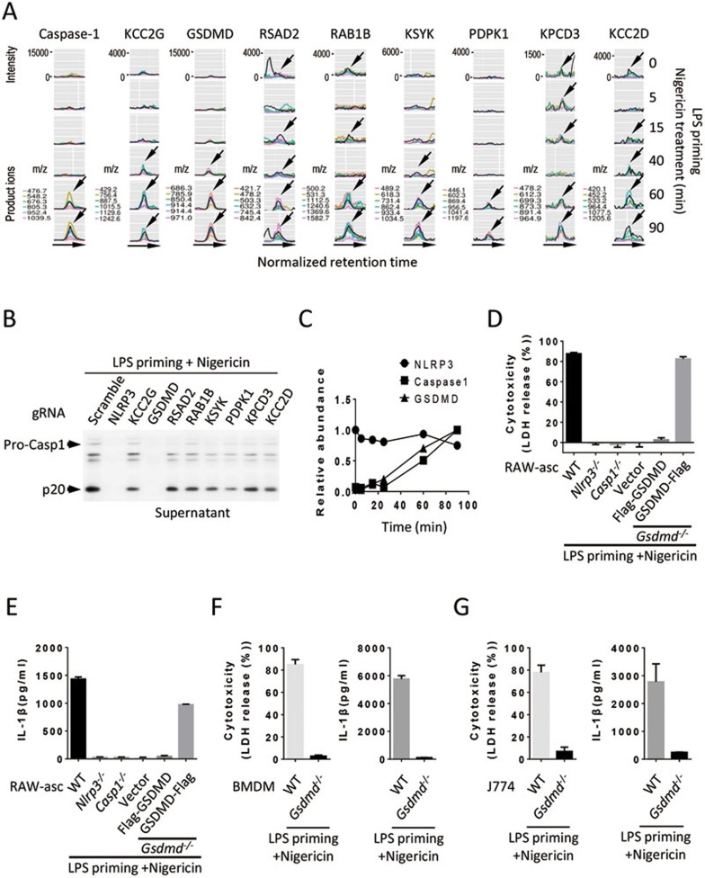
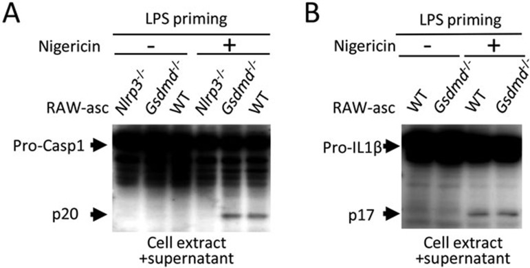
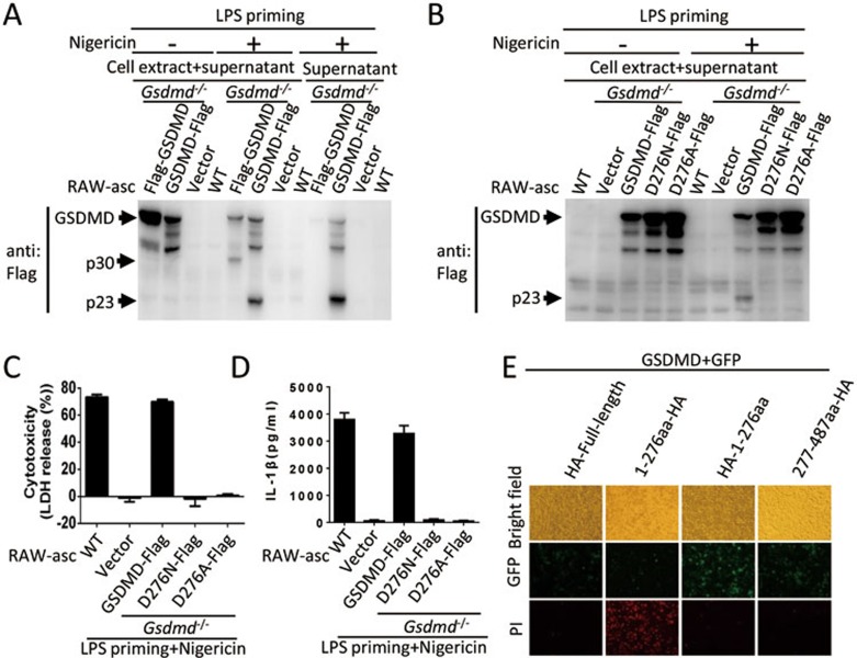
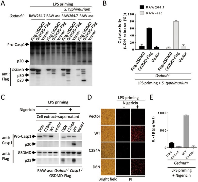
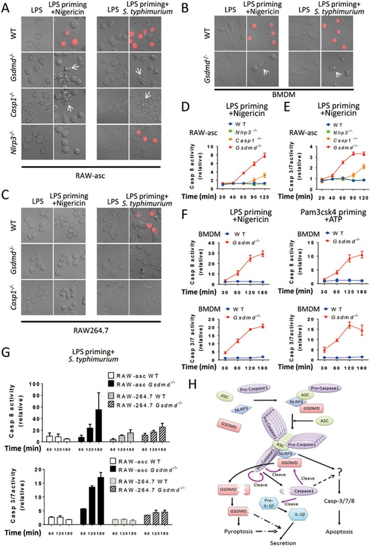
Inflammasome is an intracellular signaling complex of the innate immune system. Activation of inflammasomes promotes the secretion of interleukin 1β (IL-1β) and IL-18 and triggers pyroptosis. Caspase-1 and -11 (or -4/5 in human) in the canonical and non-canonical inflammasome pathways, respectively, are crucial for inflammasome-mediated inflammatory responses. Here we report that gasdermin D (GSDMD) is another crucial component of inflammasomes. We discovered the presence of GSDMD protein in nigericin-induced NLRP3 inflammasomes by a quantitative mass spectrometry-based analysis. Gene deletion of GSDMD demonstrated that GSDMD is required for pyroptosis and for the secretion but not proteolytic maturation of IL-1β in both canonical and non-canonical inflammasome responses. It was known that GSDMD is a substrate of caspase-1 and we showed its cleavage at the predicted site during inflammasome activation and that this cleavage was required for pyroptosis and IL-1β secretion. Expression of the N-terminal proteolytic fragment of GSDMD can trigger cell death and N-terminal modification such as tagging with Flag sequence disrupted the function of GSDMD. We also found that pro-caspase-1 is capable of processing GSDMD and ASC is not essential for GSDMD to function. Further analyses of LPS plus nigericin- or Salmonella typhimurium-treated macrophage cell lines and primary cells showed that apoptosis became apparent in Gsdmd(-/-) cells, indicating a suppression of apoptosis by pyroptosis. The induction of apoptosis required NLRP3 or other inflammasome receptors and ASC, and caspase-1 may partially contribute to the activation of apoptotic caspases in Gsdmd(-/-) cells. These data provide new insights into the molecular mechanisms of pyroptosis and reveal an unexpected interplay between apoptosis and pyroptosis.


Figures

Figure 1
Figure 1
Identification of GSDMD as a component of NLRP3 inflammasome and determination of the requirement of GSDMD in LPS plus nigericin-induced pyroptosis and IL-1β production. (A) Finding protein candidates that were recruited to NLRP3 complex by quantitative MS. NLRP3 complex was affinity-purified from NLRP3-Flag J774 cells after LPS (100 ng/ml) priming for 4 h and then nigericin (10 μM) stimulation for 0, 5, 15, 40, 60, or 90 min, digested with trypsin and subjected to high-sensitive quantitative MS analysis. MS data was analyzed with Group-DIA and peptide intensities were extracted. The results revealed nine candidate proteins whose amount time-dependently increased in NLRP3 complex. The extracted ion chromatogram (XIC) peaks of multiple product ions of one representative peptide for each of these proteins were shown. Arrows indicate the co-eluting XIC peaks of product ions of corresponding representative peptides. (B) GSDMD is involved in inflammasome activation. The genes as indicated were knocked out by CRISPR-Cas9 in RAW-asc cells. The cells were primed with LPS for 4 h and then treated with nigericin for 2 h. The release of p20 caspase-1 into the culture media was measured by immunoblotting using anti-caspase-1 antibody, which was used to assay pyroptosis. (C) Time-dependent recruitment of GSDMD and caspase-1 to NLRP3 complex. Relative abundance of NLRP3, caspase-1 and GSDMD proteins in NLRP3 immunocomplex across six time points was shown. (D) Requirement of GSDMD in pyroptosis. LDH released from RAW-asc cells with different gene deletion or gene reconstitution was measured after the cells were treated as in B. RAW-asc cells with genotype of WT, Nlrp3−/−, Caspase-1−/−(Casp1−/−), Gsdmd−/− cells reconstituted with N-terminally Flag-tagged GSDMD (Flag-GSDMD) or C-terminally Flag-tagged GSDMD (GSDMD-Flag) or a control vector were used in the experiments. (E) Requirement of GSDMD in IL-1β production. Culture supernatants of the cells described in D were analyzed by IL-1β ELISA kit. (F) Pyroptosis and IL-1β in the culture supernatants of WT and Gsdmd−/− BMDM were measured as in D, E. (G) Pyroptosis and IL-1β in the culture supernatants of WT and Gsdmd−/− J774 cells were measured as in D and E. Graphs show mean ± SD of triplicate wells and represent three independent experiments.
Figure 2
Figure 2
GSDMD is not required for proteolytic maturation of caspase-1 and IL-1β. (A) Processing of caspase-1 in Gsdmd−/− cells. Culture supernatants together with their corresponding cell extracts from WT, Nlrp3−/− and Gsdmd−/− RAW-asc cells treated as in Figure 1B were analyzed by immunoblotting with anti-caspase-1 antibody. (B) Processing of IL-1β in Gsdmd−/− cells. Culture supernatants together with their corresponding cell extracts from WT and Gsdmd−/− RAW-asc cells treated as in Figure 1B were analyzed by immunoblotting with anti-IL-1β antibody.
Figure 3
Figure 3
Cleavage of GSDMD at D276 is required for pyroptosis and IL-1β secretion and the N-terminal proteolytic fragment of GSDMD executes cell death. (A) Processing of GSDMD. WT RAW-asc cells and Gsdmd−/− RAW-asc cells reconstituted with Flag-GSDMD, GSDMD-Flag or a control vector were treated as in Figure 1B. Cell extracts together with their corresponding culture supernatants or supernatants alone were analyzed by immunoblotting with anti-Flag antibody. (B) D276 is the cutting site in GSDMD upon NLRP3 inflammasome activation. Gsdmd−/− RAW-asc cells reconstituted with the expression of GSDMD-Flag, D276N mutant of GSDMD (D276N-Flag) or D276A mutant of GSDMD (D276A-Flag) were treated as in Figure 1B. Cell extracts together with their corresponding culture supernatants were analyzed by immunoblotting with anti-Flag antibody. (C) Requirement of GSDMD cleavage in pyroptosis. LDH release was measured in the cells described in B. (D) Requirement of GSDMD cleavage in IL-1β secretion. The same as in C except that IL-1β in the culture media was measured by ELISA. (E) Pyroptosis induced by N-terminal domain of GSDMD. Green fluorescence protein (GFP) and one of the GSDMD fragments were co-expressed in 293T cells and images were taken 16 h after transfection. Graphs show mean ± SD of triplicate wells and represent three independent experiments.
Figure 4
Figure 4
GSDMD can be cleaved by pro-caspase-1. (A) GSDMD is cleaved in ASC-deficient cells in the absence of pro-caspase-1 processing. Gsdmd−/− RAW264.7 and Gsdmd−/− RAW-asc cells reconstituted with the expression of Flag-GSDMD, GSDMD-Flag or a control vector were primed with LPS for 4 h followed by S. typhimurium (100 MOI) treatment for 2 h. The cells were analyzed by immunoblotting with anti-caspase-1 and anti-Flag antibodies. (B) LDH release was measured in the cells described in A. (C) The auto-cleave-deficient pro-caspase-1 mutant can cleave GSDMD. Gsdmd and caspase-1 double knockout RAW-asc cells (Gsdmd−/− Casp1−/−) were reconstituted with GSDMD-Flag expression first and then with WT, auto-cleavage-deficient mutant D6N, and enzymatic dead mutant C284A of pro-caspase-1, respectively. The cells were treated as in Figure 1B and analyzed by immunoblotting with anti-caspase-1 and anti-Flag antibodies. (D) Pyroptosis can be mediated by auto-cleavage-deficient pro-caspase-1. The cells described in C were stained with PI and analyzed under microscope. (E) IL-1β production cannot be mediated by auto-cleavage-deficient pro-caspase-1. IL-1β in the culture media of the cells described in C was measured by ELISA. Graphs show mean ± SD of triplicate wells and represent three independent experiments.
Figure 5
Figure 5
GSDMD deletion unmasks inflammasome-induced apoptosis. (A) Apoptotic-like cells in GSDMD- or Caspase-1-deleted RAW-asc cells. WT, Gsdmd−/−, Casp1−/−, and Nlrp3−/− RAW-asc cells were used in the experiments. Images were taken after LPS priming for 4 h and then nigericin or S. typhimurium treatment for 2 h. PI was added to detect the loss of plasma membrane integrity. Arrows point to apoptotic-like cells. (B) Apoptotic-like cells in GSDMD-deleted BMDM. The same as in A except that WT and Gsdmd−/− BMDM were used and nigericin or S. typhimurium stimulation time was 1 h. (C) No apoptotic-like cells in GSDMD- or casppase-1-deleted RAW264.7 cells. The same as in A except that WT, Gsdmd−/− and Casp1−/− RAW264.7 cells were used in the experiments. (D) Apoptotic caspase-8 is activated in Gsdmd−/− and Casp1−/− RAW-asc cells after LPS plus nigericin stimulation. WT, Nlrp3−/−, Casp1−/− and Gsdmd−/− RAW-asc cells were primed with LPS for 4 h and then treated with nigericin for different time periods as indicated. The activities of caspase-8 were measured and shown. (E) Apoptotic caspase-3/7 is activated in Gsdmd−/− and Casp1−/− RAW-asc cells. The same as in D except that the activities of caspase-3/7 were measured and shown. (F) Apoptotic caspases are activated in inflammasome stimuli-treated Gsdmd−/− BMDM. WT and Gsdmd−/− BMDM were primed with LPS or Pam3csk4 (1 μg/ml) for 5 h and stimulated with nigericin or ATP (5 mM), respectively, for different periods of time as indicated. The activities of caspase-8 and caspase-3/7 were measured and shown. (G) Apoptotic caspases are not activated in ASC-deficient cells. WT and Gsdmd−/− RAW264.7 cells were primed with LPS for 4 h and stimulated with S. typhimurium for different time periods as indicated. WT and Gsdmd−/− RAW-asc cells were included for comparison. The activities of caspase-8 and caspase-3/7 were measured and shown. (H) Proposed model for pyroptosis and IL-1β production induced by NLRP3 inflammasome pathway. Graphs show mean ± SD of triplicate wells and represent three independent experiments.

References

    1. 1Martinon F, Mayor A, Tschopp J. The inflammasomes: guardians of the body. Annu Rev Immunol 2009; 27:229–265. - PubMed
    1. 2Lamkanfi M, Dixit VM. Inflammasomes and their roles in health and disease. Annu Rev Cell Dev Biol 2012; 28:137–161. - PubMed
    1. 3Fink SL, Cookson BT. Apoptosis, pyroptosis, and necrosis: mechanistic description of dead and dying eukaryotic cells. Infect Immun 2005; 73:1907–1916. - PMC - PubMed
    1. 4Netea MG, Simon A, van de Veerdonk F, et al. IL-1beta processing in host defense: beyond the inflammasomes. PLoS Pathog 2010; 6:e1000661. - PMC - PubMed
    1. 5Miao EA, Leaf IA, Treuting PM, et al. Caspase-1-induced pyroptosis is an innate immune effector mechanism against intracellular bacteria. Nat Immunol 2010; 11:1136–1142. - PMC - PubMed

Publication types