. 2016 Apr 15;17(8):693-697.
doi: 10.1002/cbic.201500632. Epub 2015 Dec 17.

Defining A-Kinase Anchoring Protein (AKAP) Specificity for the Protein Kinase A Subunit RI (PKA-RI)

Affiliations

Defining A-Kinase Anchoring Protein (AKAP) Specificity for the Protein Kinase A Subunit RI (PKA-RI)

Karolin Autenrieth et al. Chembiochem. .

Abstract

A-Kinase anchoring proteins (AKAPs) act as spatial and temporal regulators of protein kinase A (PKA) by localizing PKA along with multiple proteins into discrete signaling complexes. AKAPs interact with the PKA holoenzyme through an α-helix that docks into a groove formed on the dimerization/docking domain of PKA-R in an isoform-dependent fashion. In an effort to understand isoform selectivity at the molecular level, a library of protein-protein interaction (PPI) disruptors was designed to systematically probe the significance of an aromatic residue on the AKAP docking sequence for RI selectivity. The stapled peptide library was designed based on a high affinity, RI-selective disruptor of AKAP binding, RI-STAD-2. Phe, Trp and Leu were all found to maintain RI selectivity, whereas multiple intermediate-sized hydrophobic substitutions at this position either resulted in loss of isoform selectivity (Ile) or a reversal of selectivity (Val). As a limited number of RI-selective sequences are currently known, this study aids in our understanding of isoform selectivity and establishing parameters for discovering additional RI-selective AKAPs.

Keywords: A-kinase anchoring protein; cAMP signaling; isoforms; protein kinase A; selectivity; stapled peptides.


Figures

Figure 1
Figure 1
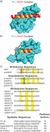
RI- versus RII-selective AKAPs. a) The RI D/D domain (blue) has a more extensive interface and engages four helical turns of the amphipathic AKAP docking helix (the dual-specific D-AKAP2 peptide is shown in red). The hydrophobic registers on the binding interface are shown in yellow. The location where the aromatic residue is positioned in an RI sequence is highlighted in orange with an arrow. b) RII D/D domain interactions with the D-AKAP2 peptide are notably different with a smaller binding interface (residues at the binding interface are shown in yellow). The position of the aromatic residue of an RI sequence is highlighted in orange with an arrow. c) AKAP docking sequences from various AKAPs demonstrates that some RI-selective sequences contain an aromatic residue in the first helical turn, but not all (i.e. PV-38). Dual-specific AKAPs such as D-AKAP1 and D-AKAP2 also lack an aromatic residue in this position but contain one on the hydrophilic face of the helix (shown in green). Unlike the RI isoform, this position (orange) does not complement a pocket on the surface of RII. Asterisks represent the positions of the non-natural olefinic amino acids in RI-STAD-2. Structures were rendered in PyMol using PDB files 3IM4 and 2HWN.
Figure 2
Figure 2
Modified RI-STAD-2 library. Using RI-STAD-2 as a template, peptide variants were generated by introducing substitutions at position 4 (orange). Aromatic, hydrophobic, polar and acidic substitutions were introduced at this site.
Figure 3
Figure 3
Log scale of KD ratios for the variants relative to RI-STAD-2. Relative KD ratios for RIα are shown in black and ratios for RIIα are shown in grey. While substitution changes were well tolerated by the RII isoform overall, the RI isoform demonstrated selectivity towards particular hydrophobic amino acids at this position.
Figure 4
Figure 4
Normalized fluorescence polarization (FP) signals for RI-STAD-2 and select variants. a) RI-STAD-2 binding curves are shown for human PKA-RIα (black squares) and PKA-RIIα (grey circles) using fluorescently labeled RI-STAD-2 b) Affinity comparison for the Leu variant (circles) and the Ile variant (squares) for the RIα and RIIα isoforms. The Ile variant binds both isoforms nearly equally, while the Leu variant displays selectivity for RIα. Further the affinity of the Leu variant to RIIα is comparable to the affinity of the Ile variant for both isoforms. c) In contrast to the binding preference for RI-STAD-2, the V variant depicts reversed selectivity with preferential binding to RIIα. d) The Asp variant had a considerable loss of affinity for both isoforms.

References

    1. Taylor SS, Yang J, Wu J, Haste NM, Radzio-Andzelm E, Anand G. Biochim Biophys Acta. 2004;1697:259–269. - PubMed
    1. Taylor SS, Zhang P, Steichen JM, Keshwani MM, Kornev AP. Biochim Biophys Acta. 2013;1834:1271–1278. - PMC - PubMed
    1. Langeberg LK, Scott JD. Nature reviews. Molecular cell biology. 2015;16:232–244. - PMC - PubMed
    1. Nygren PJ, Scott JD. Front Pharmacol. 2015;6:158. - PMC - PubMed
    1. Sarma GN, Kinderman FS, Kim C, von Daake S, Chen L, Wang BC, Taylor SS. Structure. 2010;18:155–166. - PMC - PubMed
    2. Kinderman FS, Kim C, von Daake S, Ma Y, Pham BQ, Spraggon G, Xuong NH, Jennings PA, Taylor SS. Mol Cell. 2006;24:397–408. - PMC - PubMed
    3. Gold MG, Lygren B, Dokurno P, Hoshi N, McConnachie G, Tasken K, Carlson CR, Scott JD, Barford D. Mol Cell. 2006;24:383–395. - PubMed

Publication types

MeSH terms

Substances

LinkOut - more resources