Review
. 2016 Jan;19(1):33-53.
doi: 10.1016/j.cvex.2015.08.002.

Principles of Wound Management and Wound Healing in Exotic Pets

Affiliations
Review

Principles of Wound Management and Wound Healing in Exotic Pets

Megan A Mickelson et al. Vet Clin North Am Exot Anim Pract. 2016 Jan.

Abstract

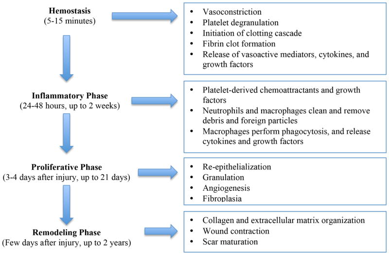
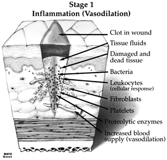
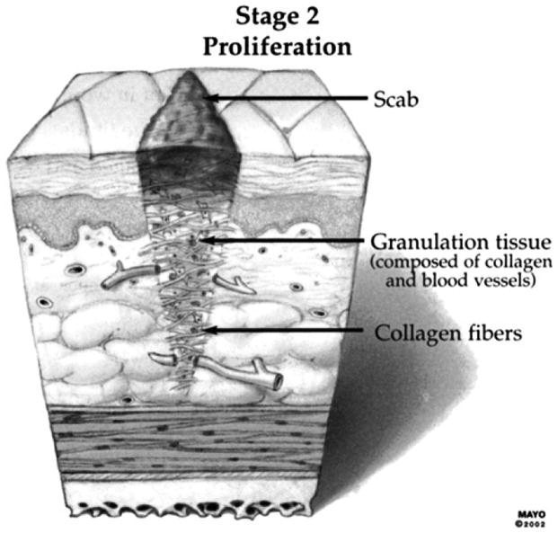
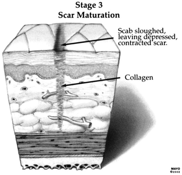
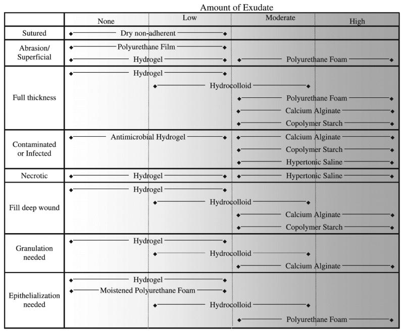
The care of wounds in exotic animal species can be a challenging endeavor. Special considerations must be made in regard to the animal's temperament and behavior, unique anatomy and small size, and tendency toward secondary stress-related health problems. It is important to assess the entire patient with adequate systemic evaluation and consideration of proper nutrition and husbandry, which could ultimately affect wound healing. This article summarizes the general phases of wound healing, factors that affect healing, and principles of wound management. Emphasis is placed on novel methods of treating wounds and species differences in wound management and healing.

Keywords: Topical wound therapy; Wound dressings; Wound healing; Wound management; Wound products.


Figures

Figure 1
Figure 1
Stages of wound healing. (Adapted from Ozturk F, Ermertcan AT. Wound healing: a new approach to the topical wound care. Cutan Ocul Toxicol 2011;30:92–99, with permission.)
Figure 2
Figure 2
Stage 1 of wound healing: Inflammation (vasodilation) (From Sherris DA, Kern EB. Essential surgical skills. 2nd edition. Philadelphia: WB Saunders; 2004. p. 13. By permission of Mayo Foundation for Medical Education and Research. All rights reserved.)
Figure 3
Figure 3
Stage 2 of wound healing: Proliferation. (From Sherris DA, Kern EB. Essential surgical skills. 2nd edition. Philadelphia: WB Saunders; 2004. p. 13. By permission of Mayo Foundation for Medical Education and Research. All rights reserved.)
Figure 4
Figure 4
Stage 3 of wound healing: Scar maturation. (From Sherris DA, Kern EB. Essential surgical skills. 2nd edition. Philadelphia: WB Saunders; 2004. p. 13. By permission of Mayo Foundation for Medical Education and Research. All rights reserved.)
Figure 5
Figure 5
Skin laceration in a rabbit. A) Initial presentation of the wound, within one hour after infliction due to con-specific trauma. B) The wound was lavaged the skin edges trimmed and skin staples were used to perform primary wound closure. The wound healed without complications.
Figure 6
Figure 6
Chronic wound dorsal to the tail base in a pained turtle (Chrysemys picta) secondary to marginal scute malformation allowing for con-specific trauma in an overcrowded enclosure. A) Initial presentation. B) The same wound following surgical debridement. Note the healthy granulation tissue
Figure 7
Figure 7
Lavage of a pododermatitis wound following surgical debridement in a peregrine falcon (Falco peregrinus).
Figure 8
Figure 8
Dressing of a carapacial wound in a common snapping turtle (Chelydra serpentina). A hydrogel sheet is applied to the wound and secured in place by a waterproof transparent film dressing. The animal was allowed full access to water, because the dressing was waterproof.
Figure 9
Figure 9
A) Carapacial fracture repair followed by application silver sulfadiazine cream (B) to the fracture site following reduction, in order to prevent contamination in a Blanding’s turtle (Emys blandingii).
Figure 10
Figure 10
Large full-thickness wound over the dorsum in a blue and gold macaw (Ara ararauna). A) A hydrogel sheet is placed on the wound as the primary dressing. Note the loose suture loops placed in the periphery of the wound in order to facilitate the tie-over-bandage. B) Gauze has been used as the secondary bandage layer. Umbilical tape is used to create a tie-over bandage. For additional support a waterproof transparent film dressing was applied.
Figure 11
Figure 11
A) Full-thickness wound of the distal tarsometatarsus of a wattled crane (Bugeranus carunculatus). B) Application of a commercial honey dressing sheet to the same wound. A waterproof transparent film dressing is applied as the secondary layer in order to secure the honey dressing in place. An elastic bandage was applied as the tertiary layer (not shown).
Figure 12
Figure 12
Multiple Carapacial fractures and bone loss in a common snapping turtle (Chelydra serpentine). A) Initial presentation. B) Following surgical debridement and stabilization of the fractures, sterile gauze strips soaked in raw honey were applied to the soft tissue wounds. C) A waterproof transparent film dressing is applied as the outer layer and the edges are secured with medical tape.
Figure 13
Figure 13
Guidelines for selection of primary wound dressings based on wound type and amount of wound exudate. (From Campbell BG. Dressings, Bandages, and splints for wound management in dogs and cats. Vet Clin Small Anim 2006;36:759–91, with permission.)

References

    1. Portou MJ, Baker D, Abraham D, et al. The innate immune system, toll-like receptors and dermal wound healing: A review. Vascul Pharmacol. 2015 Epub ahead of print. - PubMed
    1. Ozturk F, Ermertcan AT. Wound healing: a new approach to the topical wound care. Cutan Ocul Toxicol. 2011;30:92–99. - PubMed
    1. Pazyar N, Yaghoobi R, Rafiee E, et al. Skin wound healing and phytomedicine: a review. Skin Pharmacol Physiol. 2014;27:303–310. - PubMed
    1. Cornell K. Wound Healing. In: Tobias KM, Johnston SA, editors. Veterinary Surgery: Small Animal. 1. Vol. 1. St. Louis: Saunders; 2012. pp. 125–134.
    1. Fossum, Hedlund CS. Small Animal Surgery. 3. St. Louis: Mosby, Inc; 2007. Surgery of the Integumentary System; pp. 159–228.

MeSH terms

Substances