. 2016 Feb 1;25(3):472-83.
doi: 10.1093/hmg/ddv486. Epub 2015 Nov 26.

Functional characterization of a human POU1F1 mutation associated with isolated growth hormone deficiency: a novel etiology for IGHD

Affiliations

Functional characterization of a human POU1F1 mutation associated with isolated growth hormone deficiency: a novel etiology for IGHD

Marie-Laure Sobrier et al. Hum Mol Genet. .

Abstract

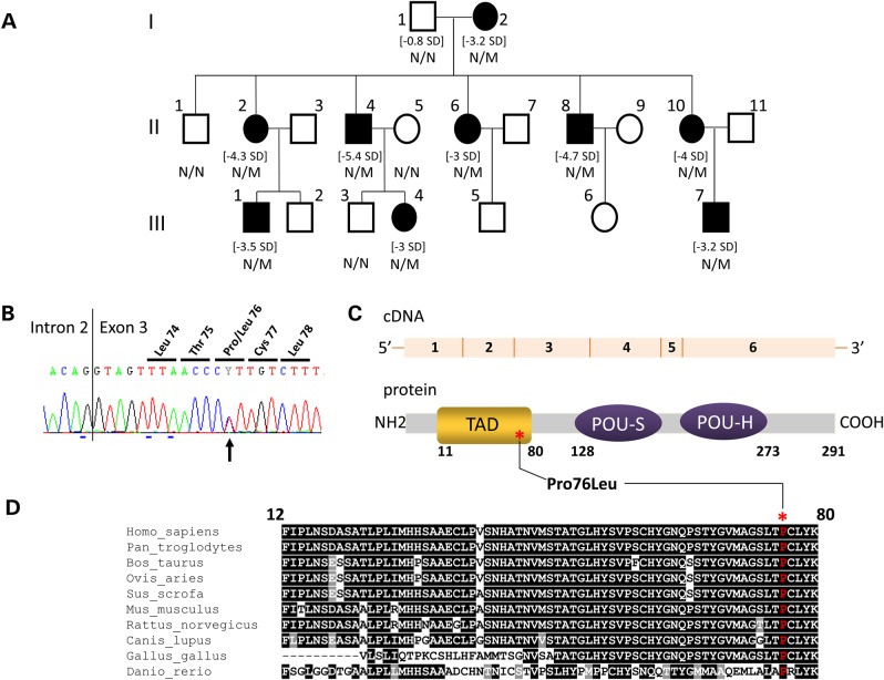
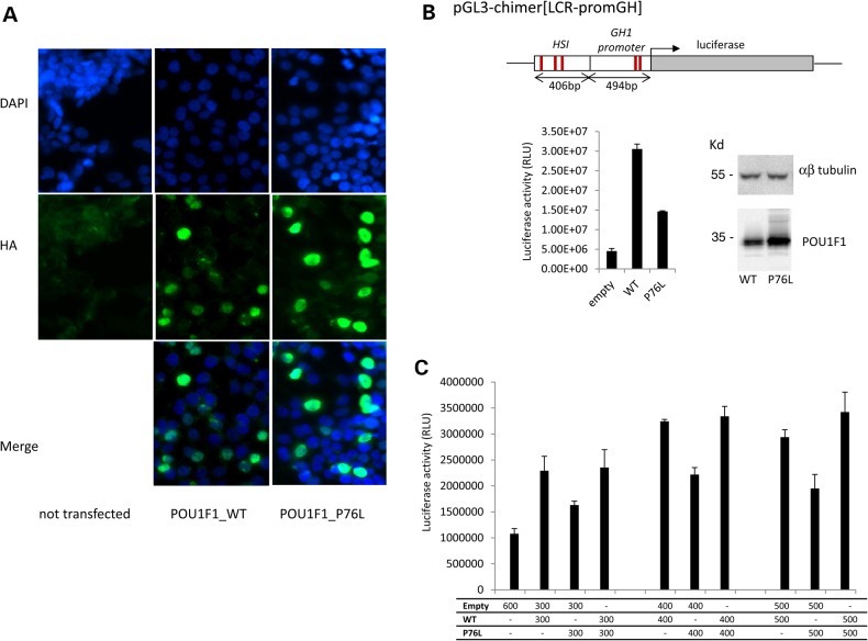
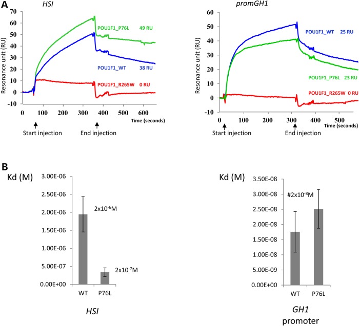
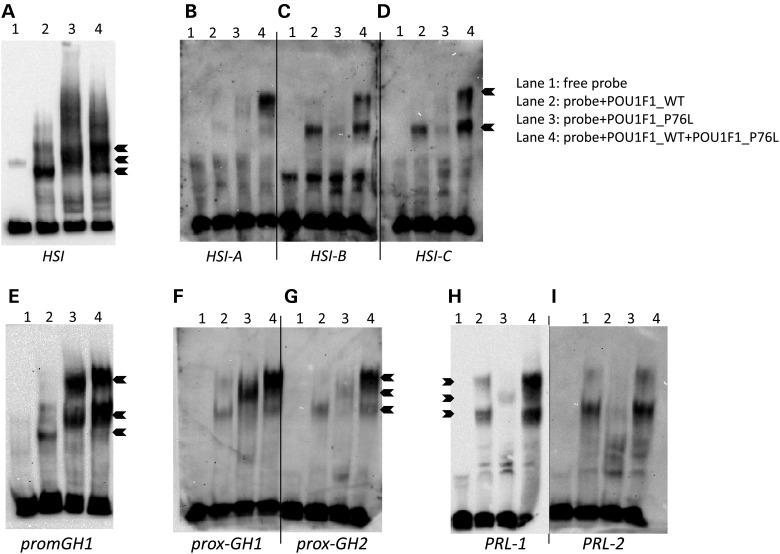
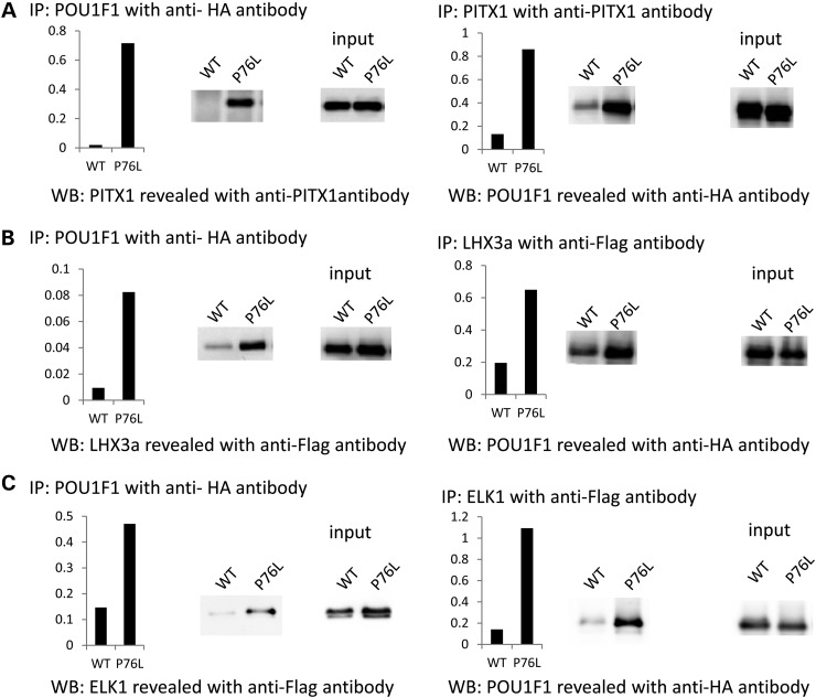
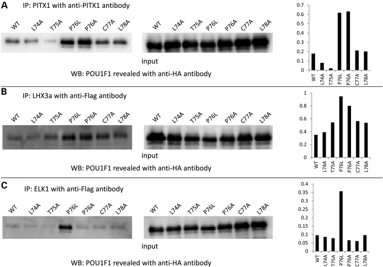
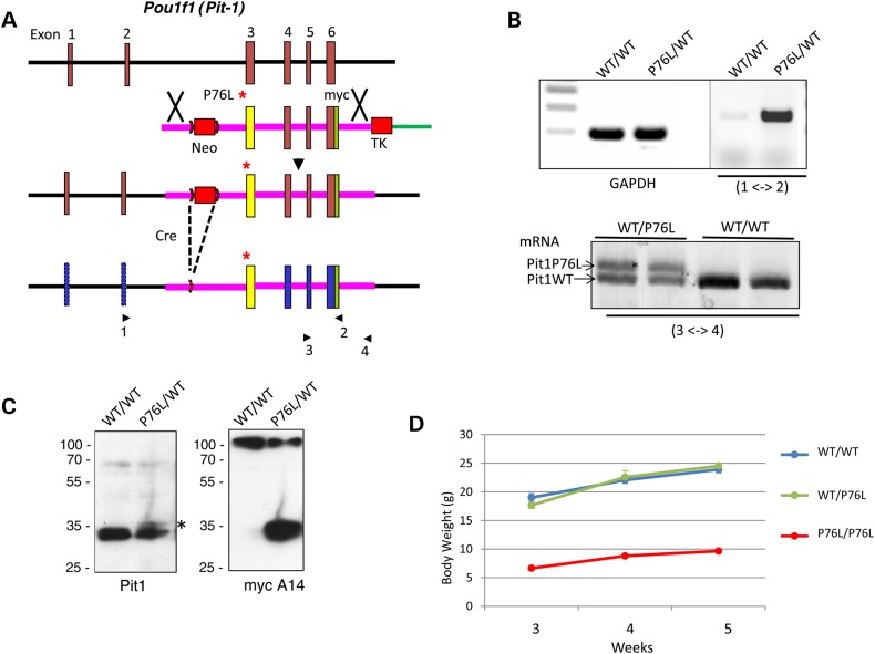
POU1F1, a pituitary-specific POU-homeo domain transcription factor, plays an essential role in the specification of the somatotroph, lactotroph and thyrotroph lineages and in the activation of GH1, PRL and TSHβ transcription. Individuals with mutations in POU1F1 present with combined deficiency of GH, PRL and TSH. Here, we identified a heterozygous missense mutation with evidence of pathogenicity, at the POU1F1 locus, in a large family in which an isolated growth hormone deficiency segregates as an autosomal dominant trait. The corresponding p.Pro76Leu mutation maps to a conserved site within the POU1F1 transactivation domain. Bandshift assays revealed that the mutation alters wild-type POU1F1 binding to cognate sites within the hGH-LCR and hGH1 promoter, but not to sites within the PRL promoter, and it selectively increases binding affinity to sites within the hGH-LCR. Co-immunoprecipitation studies reveal that this substitution enhances interactions of POU1F1 with three of its cofactors, PITX1, LHX3a and ELK1, and that residue 76 plays a critical role in these interactions. The insertion of the mutation at the mouse Pou1f1 locus results in a dramatic loss of protein expression despite normal mRNA concentrations. Mice heterozygous for the p.Pro76Leu mutation were phenotypically normal while homozygotes demonstrated a dwarf phenotype. Overall, this study unveils the involvement of POU1F1 in dominantly inherited isolated GH deficiency and demonstrates a significant impact of the Pro76Leu mutation on DNA-binding activities, alterations in transactivating functions and interactions with cofactors. Our data further highlight difficulties in modeling human genetic disorders in the mouse despite apparent conservation of gene expression pathways and physiologic functions.


Figures

Figure 1.
Figure 1.
Identification of a POU1F1 mutation segregating with a short stature phenotype and low serum GH levels in a three generation kindred. (A) Genealogical tree of the IGHD family. Squares: males; circles: females; filled black symbols: IGHD patients. For subjects with growth retardation, their height SD is indicated between brackets. The genotype (N/N: normal, N/M: heterozygous) is indicated under each tested individual. (B) Electrophoregram of the portion of Exon 3 showing (black arrow) the heterozygous c.227C > T transition leading to p.Pro76Leu mutation. The vertical line represents the intron–exon junction. (C) A schematic representation of the POU1F1 cDNA and POU1F1 protein: six exons encoding the 291 amino acids protein consisting of two main domains, the TAD (orange) in which the variation has been identified (noted with an asterisk) and the POU-S and POU-H domains (homeodomain, purple). (D) Evolutionary conservation of proline 76 (noted above with an asterisk): interspecies similarity (shown in one-letter code) of the TAD domain of POU1F1 aligned with sequences of the TAD domain found in nine other vertebrates species: black underlined, total conservation; gray underlined, conservative amino acid substitutions; not underlined, amino acid not conserved and indexed in a different group.
Figure 2.
Figure 2.
Conserved nuclear localization and diminished transcriptional activity of the mutant POU1F1 protein. (A) Subcellular localization of POU1F1_WT and POU1F1_P76L in HEK293T cells transfected with the corresponding HA-tagged expression plasmids. Forty-eight hours after transfection, cells were immunostained with mouse anti-HA antibody (1/1000) then Alexa488 (goat anti-mouse 1/2000). Nuclei are stained in blue by DAPI. The two proteins (WT and mutated) were both localized in the nucleus. A control with no transfected cells is also shown. (B) Impact of the Pro76Leu mutation on the transcriptional capability of POU1F1. HEK293T cells were co-transfected with pcDNA3-POU1F1_WT-HA or pcDNA3-POU1F1_P76L-HA in combination with a luciferase reporter ORF under control of the HSI enhancer segment of the hGH LCR linked directly to the intact hGH promoter pGL3-chimer[LCR-promGH]. The previously defined POU1F1-binding sites in HSI and in the hGH promoter are indicated by the red lines. POU1F1 protein expression from the expression vectors was monitored by western blot with anti-POU1F1 polyclonal antibody relative to αβ-tubulin level (top). Luciferase activity represents the means ± SD of triplicate assays; a representative experiment of three experiments. (C) The assessment of a potential dominant-negative effect of the P76L mutation over the WT protein. Co-transfection of the HSI/hGH/Luc reporter with the POU1F1_WT plasmid, POU1F1_P76L plasmid in increasing amounts up to a saturation response or with a 1:1 mixture of the two plasmids.
Figure 3.
Figure 3.
POU1F1 binding to DNA studied by SPR. (A) Sensorgrams. Biotinylated DNAs (HSI and promGH1) were loaded on a streptavidin chip and binding of 50 nm of purified WT (blue) or P76L (green) or R265W (red) POU1F1 proteins to the two DNA targets was assessed. The recorded RUs for each protein were noted. (B) Kinetics of POU1F1 binding the DNA sites: 0, 10, 20, 30, 40 and 50 nm of each purified protein were successively loaded on the streptavidin chips on which biotinylated DNA POU1F1 targets have been anchored. After data treatment using the BIAevaluation software 4.1, the association and dissociation constants corresponding to a Kd value in molar (in m) were determined for WT and P76L-POU1F1 proteins on each target sequence and represented by histograms: on the HSI (left graph) and on the hGH1 promoter (right graph). Each study was performed in triplicate.
Figure 4.
Figure 4.
Analysis of POU1F1 binding to DNA by electrophoretic mobility shift assays. (A) Twenty fmoles of biotinylated DNA containing a 212 bp segment of HSI encompassing all three POU1F1-binding sites (A, B and C) were incubated with 200 ng of purified WT (Lane 2) or mutant P76L POU1F1 (Lane 3) protein or with a 1:1 mixture (100 ng of each) of the two proteins (Lane 4). Lane 1 contains the biotinylated DNA target in the absence of added protein. (BD) Binding to each individual POU1F1-binding site within HSI: 37 bp including one site HSI-A, B, 37 bp HSI-B, C, 38 bp HSI-C, D. (E) Binding to a 70 bp segment of the GH1 promoter encompassing the two POU1F1-binding sites. (FG) Binding to each of the two POU1F1-binding sites in the hGH1 promoter: 32 bp fragment encompassing prox-GH1 (F) or the 30 bp fragment encompassing prox-GH2 (G). (H and I) Binding to each of the two POU1F1-binding sites in the human PRL gene promoter: 29 bp fragments containing either PRL-1 (H) or PRL-2 (I).
Figure 5.
Figure 5.
Co-immunoprecipitation of WT and P76L POU1F1 proteins with three cofactors. HEK293T cells were co-transfected with plasmids pcDNA4-POU1F1-HA expressing POU1F1_WT or P76L (lanes labeled WT and P76L, respectively) and with each of the following three expression vectors: pcDNA3-PITX1 (A), pcDNA3-LHX3a-Flag (B) and pcDNA3-ELK1-Flag (C). Nuclear extracts were co-immunoprecipitated with an anti-HA antibody, and the western blots were revealed with an anti-PITX1 antibody for PITX1 and an anti-Flag antibody for LHX3 and ELK1 (left panels). The same nuclear extracts were co-immunoprecipitated with an anti-PITX1 antibody for PITX1 and an anti-Flag antibody for LHX3 and ELK1, and westerns blots revealed with the anti-HA antibody (right panels); IP: immunoprecipitation, WB: western blots. Quantification of the complex formed, represented by histograms, was calculated as the ratio of protein immunoprecipitated to protein expressed (input).
Figure 6.
Figure 6.
Co-immunoprecipitation POU1F1 with three cofactors. Interactions are compared among the WT POU1F1 and a series of derived alanine substitutions surrounding proline 76 site. HEK293T cells were co-transfected with plasmids pcDNA4-POU1F1-HA expressing POU1F1_WT or L74A, T75A, P76L, P76A, C77A, L78A and pcDNA3-PITX1 or pcDNA3-LHX3a-Flag or pcDNA3-ELK1-Flag. Co-immunoprecipitations were performed with an anti-PITX1 antibody (A) or Flag antibodies for LHX3a (B) and ELK1 (C) on nuclear extracts samples. Western blots were generated using an anti-HA antibody. Quantification of the complex formed, represented by histograms (right side), was evaluated as the ratio of protein immunoprecipitated to protein expressed (input).
Figure 7.
Figure 7.
Introduction of the P76L mutation into the Pou1f1 locus in the mouse genome. (A) Homologous recombination at the Pou1f1 locus in the mouse genome. The native-Pou1f1 locus is shown on the top and the targeting vector used to insert the P76L mutation into the locus is shown below. The initial recombination product is displayed on the third line and the final locus after deletion of the NeoR cassette via Cre/Lox recombination is displayed at the bottom. The position of the P76L mutation is denoted by the asterisk, the dual selection cassettes, NeoR and thymidine kinase, are represented by the labeled red rectangles. Lox sites are indicated by the arrowhead and the positions of the primers used for selective detection of the wt and mutant Pou1f1 mRNAs are indicated and numbered below the diagram. (B) mRNA expression from the PitP76L locus in the mouse pituitary. Top: Pou1f1 mRNA expression from the P76L locus was specifically detected in a mouse heterozygous for the mutant allele by an RT/PCR assay using a set of primers positioned at Exon 2 (Primer 1) and within the myc epitope segment in Exon 6 (specific to the mutant allele) (Primer 2). Bottom: the relative levels of the mRNA expression from the wt and the mutant locus were directly compared in a mouse heterozygous for the mutant allele (wt/P76L) and a wt mouse (wt/wt) by an RT/PCR analysis using the Exon 5 primer (Primer 3) and a primer within the 3′ untranslated region (Primer 4) in Exon 4, bracketing the myc epitope tag specific to the mutant locus. (C) Protein expression from the P76L locus in the mouse pituitary. Western blot of pituitary extracts from wt/wt and wt/P76L mice were probed with antibodies to mouse Pou1f1 (left blot) and with an antibody to the myc epitope tag (specific to the mutant locus) (right blot). The overall level of Pou1f1_WT protein in the WT/P76L heterozygote pituitary is approximately half of that in the WT/WT mouse. A faint band at the position of the mutant Pit1 protein (asterisk) is migrating at the position predicted from the Pit-1 protein containing the Myc epitope tag. The expression of the P76L Pou1f1 protein was directly verified by re-probing the western with an antibody to the myc epitope. (D) Growth curves of WT/WT, WT/P76L and P76L/P76L mice. The body weights are shown in the Y-axis and the genotypes are indicted for each curve. The analyses of male mice are shown; a parallel analysis of females gave an identical result (not shown).

References

    1. Rizzoti K. (2015) Genetic regulation of murine pituitary development. J. Mol. Endocrinol., 54, R55–R73. - PMC - PubMed
    1. Andersen B., Rosenfeld M.G. (2001) POU domain factors in the neuroendocrine system: lessons from developmental biology provide insights into human disease. Endocr. Rev., 22, 2–35. - PubMed
    1. Ho Y., Cooke N.E., Liebhaber S.A. (2015) An autoregulatory pathway establishes the definitive chromatin conformation at the pit-1 locus. Mol. Cell Biol., 35, 1523–1532. - PMC - PubMed
    1. Ingraham H.A., Flynn S.E., Voss J.W., Albert V.R., Kapiloff M.S., Wilson L., Rosenfeld M.G. (1990) The POU-specific domain of Pit-1 is essential for sequence-specific, high affinity DNA binding and DNA-dependent Pit-1-Pit-1 interactions. Cell, 61, 1021–1033. - PubMed
    1. Andersen B., Rosenfeld M.G. (1994) Pit-1 determines cell types during development of the anterior pituitary gland. A model for transcriptional regulation of cell phenotypes in mammalian organogenesis. J. Biol. Chem., 269, 29335–29338. - PubMed

Publication types

MeSH terms