. 2016 Apr;75(4):721-9.
doi: 10.1136/annrheumdis-2015-208093. Epub 2015 Nov 26.

Identification of a novel chemokine-dependent molecular mechanism underlying rheumatoid arthritis-associated autoantibody-mediated bone loss

Affiliations

Identification of a novel chemokine-dependent molecular mechanism underlying rheumatoid arthritis-associated autoantibody-mediated bone loss

Akilan Krishnamurthy et al. Ann Rheum Dis. 2016 Apr.

Erratum in

Abstract

Objectives: Rheumatoid arthritis (RA)-specific anti-citrullinated protein/peptide antibodies (ACPAs) appear before disease onset and are associated with bone destruction. We aimed to dissect the role of ACPAs in osteoclast (OC) activation and to identify key cellular mediators in this process.

Methods: Polyclonal ACPA were isolated from the synovial fluid (SF) and peripheral blood of patients with RA. Monoclonal ACPAs were isolated from single SF B-cells of patients with RA. OCs were developed from blood cell precursors with or without ACPAs. We analysed expression of citrullinated targets and peptidylarginine deiminases (PAD) enzymes by immunohistochemistry and cell supernatants by cytometric bead array. The effect of an anti-interleukin (IL)-8 neutralising antibody and a pan-PAD inhibitor was tested in the OC cultures. Monoclonal ACPAs were injected into mice and bone structure was analysed by micro-CT before and after CXCR1/2 blocking with reparixin.

Results: Protein citrullination by PADs is essential for OC differentiation. Polyclonal ACPAs enhance OC differentiation through a PAD-dependent IL-8-mediated autocrine loop that is completely abolished by IL-8 neutralisation. Some, but not all, human monoclonal ACPAs derived from single SF B-cells of patients with RA and exhibiting distinct epitope specificities promote OC differentiation in cell cultures. Transfer of the monoclonal ACPAs into mice induced bone loss that was completely reversed by the IL-8 antagonist reparixin.

Conclusions: We provide novel insights into the key role of citrullination and PAD enzymes during OC differentiation and ACPA-induced OC activation. Our findings suggest that IL8-dependent OC activation may constitute an early event in the initiation of the joint specific inflammation in ACPA-positive RA.

Keywords: Ant-CCP; Autoantibodies; Early Rheumatoid Arthritis.


Figures

Figure 1
Figure 1
Polyclonal (anti-CCP2 affinity-purified) and monoclonal (single B-cell-derived) anti-citrullinated protein/peptide antibodies (ACPAs) induce osteoclast (OC) activation and bone resorption. (A) Tartrate-resistant acid phosphatase (TRAP) staining of mature OCs obtained from Mφ derived from CD14-positive monocytes of healthy individuals and cultured in the presence of either non-ACPA flow-through IgGs or ACPA IgGs (ACPA) at a concentration of 0.1 µg/mL (original magnification 200×). The graph represents the fold increase in OC (TRAP-positive cells with ≥3 nuclei) numbers and fold increase in resorption areas. The values represent the mean±SEM of three independent experiments. (B) TRAP staining of mature OCs and microscopic visualisation of calcium phosphate resorption areas in the presence of four monoclonal ACPAs (ie, B02, D10, B09 and C07) and one control anti-tetanus monoclonal antibody (ie, E02) at a concentration of 1 µg/mL. The graphs represent fold increases in OC (TRAP-positive cells with ≥3 nuclei) numbers and fold increases in resorption area. The values represent the mean±SEM of four independent experiments. (C) Graphs represent the mean±SEM of dose titrations experiments of stimulatory B02 and non-stimulatory B09 ACPA in OC (number of TRAP-positive cells with ≥3 nuclei) and bone resorption assay (resorption area in %). (D) TRAP staining of mature OCs and microscopic visualisation of calcium phosphate resorption area in the presence of Fab fragments of D10, B02 and E02 antibodies (1 µg/mL). (N=4). The graphs represent fold increases in OC (TRAP-positive cells with ≥3 nuclei) numbers and fold increases in resorption area. The values represent the mean±SEM of four independent experiments. *p<0.05.
Figure 2
Figure 2
Expression of citrullinated targets and peptidylarginine deiminases (PAD) enzymes during different stages of osteoclast (OC) differentiation. (A) immunohistochemistry images showing brown 3,3-diaminobenzidene (DAB) staining of citrullinated targets in different stages of differentiation from CD14-positive monocyte precursors to Mφ and mature OCs. Slides were stained with murinised monoclonal anti-citrullinated protein/peptide antibodies (ACPAs) (mB02, mD10, mC07) and a monoclonal control antibody (mE02) and counterstained with haematoxylin (original magnification 500× for CD14-positive monocytes and mature OCs and 250× for the intermediate stages). (B) Confocal microscopy images showing red fluorescence staining with monoclonal murinised ACPAs (mE02, mB02, mD10, mC07) and blue 4′,6-diamidino-2-phenylindole (DAPI) nuclear staining in mature OC. (C) Confocal microscopy images showing red fluorescence staining with polyclonal ACPAs, green fluorescence with anti CD68 antibody and blue nuclear staining with DAPI in mature OCs. (D) Immunohistochemistry images showing brown DAB staining of PAD2 and PAD4 expression in different stages of differentiation from CD-14-positive monocyte precursors to Mφ and mature OCs. Slides were counterstained with haematoxylin (original magnification 250×). (E) PAD activity was measured using an antibody-based assay by adding Mφ and OC cell lysates to arginine-coated plates, followed by ELISA measurement of the amounts of deiminated arginine. The graph represents the PAD enzyme activity expressed in mU/mg protein. The values represent the mean±SEM of two independent experiments. (F) Immunohistochemistry images showing brown DAB staining of citrullinated targets in mature OCs with or without incubation with a PAD inhibitor (Cl-amidine) added from the beginning of the cultures. Slides were stained with murinised monoclonal ACPAs (mB02) and a monoclonal control antibody (mE02) and counterstained with haematoxylin (original magnification 250×).
Figure 3
Figure 3
Peptidylarginine deiminases (PAD) enzymes are essential for osteoclastogenesis and the anti-citrullinated protein/peptide antibody (ACPA)-mediated effect. (A) PAD inhibition (PADi, Cl-amidine) dose-dependently inhibited osteoclast (OC) differentiation and maturation without any cytotoxic effect. The graphs represent fold decreases in OC (tartrate-resistant acid phosphatase (TRAP)-positive cells with ≥3 nuclei) numbers and fold increases in LDH release in the culture supernatants. The values represent the mean±SEM. (B) PADi does not affect either SF migration or survival. The graphs represent fold increases in the migration index of synovial fibroblast and LDH release in the culture supernatants. The values represent the mean±SEM. (C) the addition of PADi from the beginning of the OC cultures prevented ACPA-induced OC activation and calcium phosphate resorption. The graphs represent fold increases in OC (TRAP-positive cells with ≥3 nuclei) numbers. The values represent the mean±SEM of three independent experiments. Images represent the resorption area by OCs (original magnification 40×). (D) Dose titration of PADi showing that early PAD inhibition (at the initiation of the OC culture) with doses as low as 0.2 μM PADi inhibits ACPA-mediated osteoclastogenesis but no longer the unstimulated differentiation of OCs. The graphs represent fold decreases in OC (TRAP-positive cells with ≥3 nuclei) numbers. The values represent the mean±SEM. (E) Late PAD inhibition (3 days before ending the OC cultures) inhibited ACPA-mediated osteoclastogenesis but not the unstimulated differentiation of OCs. The graphs represent fold increases in OC (TRAP-positive cells with ≥3 nuclei) numbers. The values represent the mean±SEM. *p<0.05.
Figure 4
Figure 4
Interleukin (IL)-8 is an essential mediator of anti-citrullinated protein/peptide antibody (ACPA)-driven osteoclastogenesis. (A) cytometric bead array showed that ACPA, but not control IgGs, increased IL-8 release in the culture supernatants of mature osteoclasts (OCs). The values represent the mean±SEM of three independent experiments. (B) Cytometric bead array showed high levels of IL-8 in Mφ-derived OC cultures at early time points during their maturation, which further increased over time. ACPA, but not control IgGs, additionally increased IL-8 release in the culture supernatants at all time points tested. The graph shows a representative time kinetic variation in IL-8 concentrations in cell culture supernatants from one of the three tested donors. The values represent the mean±SEM. (C) Exogenous added IL-8 increases osteoclastogenesis in a dose-dependent manner. The values represent the mean±SEM. (D) Neutralising anti-IL-8 antibodies inhibited Mφ-derived OCs maturation dose dependently. The graphs represent fold decreases in OC (tartrate-resistant acid phosphatase (TRAP)-positive cells with ≥3 nuclei) numbers. The values represent the mean±SEM of three independent experiments. (E) Anti-IL-8 neutralising antibodies completely abolished the effect of ACPAs at doses as low as 1 μg/mL. The graphs represent fold increases in OC (TRAP-positive cells with ≥3 nuclei) numbers. The values represent the mean±SEM of three independent experiments. (F) Both early (first three days of culture) and late (last three days of the culture) addition of anti-IL-8 neutralising antibodies (1 μg/mL) completely abolished the effect of ACPAs. The graphs represent fold increases in OC (TRAP-positive cells with ≥3 nuclei) numbers. The values represent the mean±SE. (G) Anti-IL-8 neutralising antibodies but not an antibody against tumour necrosis factor (TNF)-α (adalimumab) abolished the effect of ACPAs at concentrations as high as 10 μg/mL. The graphs represent fold increases in OC (TRAP-positive cells with ≥3 nuclei) numbers. The values represent the mean±SEM of three independent experiments. *p<0.05.
Figure 5
Figure 5
Anti-citrullinated protein/peptide antibodies (ACPAs) induce systemic bone loss in vivo that is reversed by interleukin (IL)-8 inhibition. Representative 2D micro-CT images of the tibial metaphysis of control mice (A, n=7) and mice that were injected with ACPAs in the absence (B, n=9) or presence of reparixin (C, n=9). (B) Graphs showing quantitative evaluation of the trabecular bone mineral density (BMD, D), trabecular number (E), bone volume fraction (bone volume/tissue volume, F) and the cortical tissue mineral density (TMD, G). The values represent the mean±SEM. *p<0.05.
Figure 6
Figure 6
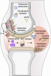
Schematic illustration of the peptidylarginine deiminases (PAD)-dependent differentiation and maturation of osteoclasts (OCs), allowing initial OC targeting by anti-citrullinated protein/peptide antibodies (ACPAs) with consecutive interleukin (IL)-8 release. OC precursors (OCPs) are present in the bone marrow and can develop into mature OCs. During the differentiation and activation of OCP, a gradual increase in cell citrullination occurred as a consequence of increased PAD activity in a calcium-rich microenvironment. ACPAs present in the circulation can reach and bind to maturing OCPs in the bone marrow, leading to an increase in OC activity with consecutive bone resorption through an IL-8-dependent autocrine loop. In a second step, IL-8 will reach the joint and initiate the chemoattraction and migration of inflammatory cells in particular neutrophils. Neutrophil extracellular traps are released by these neutrophils in the presence of ACPAs, which further contributes to the initiation of joint inflammation with the local accumulation of other inflammatory cells (such as macrophages) and activation of synovial fibroblasts, resulting in synovial membrane inflammation. NET, neutrophil extracellular traps.

Comment in

References

    1. Klareskog L, Catrina AI, Paget S. Rheumatoid arthritis. Lancet 2009;373:659–72. 10.1016/S0140-6736(09)60008-8 - DOI - PubMed
    1. Rantapää-Dahlqvist S, de Jong BA, Berglin E, et al. . Antibodies against cyclic citrullinated peptide and IgA rheumatoid factor predict the development of rheumatoid arthritis. Arthritis Rheum 2003;48:2741–9. 10.1002/art.11223 - DOI - PubMed
    1. de Hair MJ, van de Sande MG, Ramwadhdoebe TH, et al. . Features of the synovium of individuals at risk of developing rheumatoid arthritis: implications for understanding preclinical rheumatoid arthritis. Arthritis Rheumatol 2014;66:513–22. 10.1002/art.38273 - DOI - PMC - PubMed
    1. van der Helm-van Mil AH, Verpoort KN, Breedveld FC, et al. . Antibodies to citrullinated proteins and differences in clinical progression of rheumatoid arthritis. Arthritis Res Ther 2005;7:R949–958. 10.1186/ar1767 - DOI - PMC - PubMed
    1. Syversen SW, Goll GL, van der Heijde D, et al. . Prediction of radiographic progression in rheumatoid arthritis and the role of antibodies against mutated citrullinated vimentin: results from a 10-year prospective study. Ann Rheum Dis 2010;69:345–51. 10.1136/ard.2009.113092 - DOI - PubMed

Publication types

MeSH terms