. 2016 Mar 1;39(3):511-21.
doi: 10.5665/sleep.5516.

Upper Airway Collapsibility (Pcrit) and Pharyngeal Dilator Muscle Activity are Sleep Stage Dependent

Affiliations

Upper Airway Collapsibility (Pcrit) and Pharyngeal Dilator Muscle Activity are Sleep Stage Dependent

Jayne C Carberry et al. Sleep. .

Abstract

Study objectives: An anatomically narrow/highly collapsible upper airway is the main cause of obstructive sleep apnea (OSA). Upper airway muscle activity contributes to airway patency and, like apnea severity, can be sleep stage dependent. Conversely, existing data derived from a small number of participants suggest that upper airway collapsibility, measured by the passive pharyngeal critical closing pressure (Pcrit) technique, is not sleep stage dependent. This study aimed to determine the effect of sleep stage on Pcrit and upper airway muscle activity in a larger cohort than previously tested.

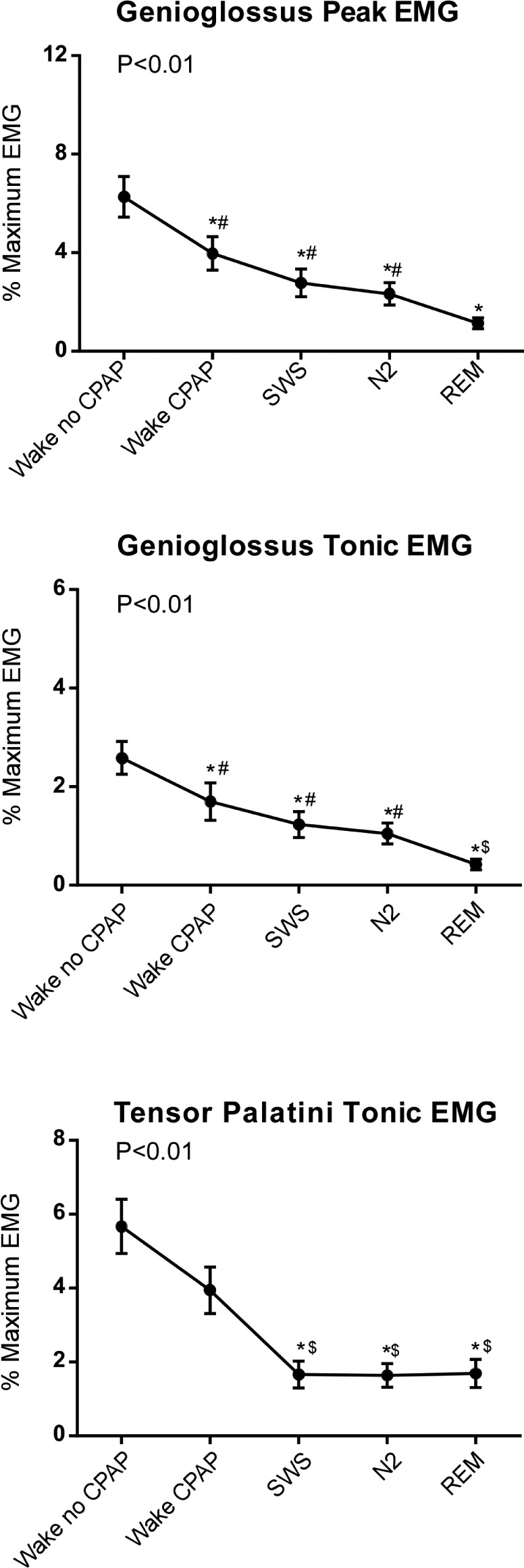
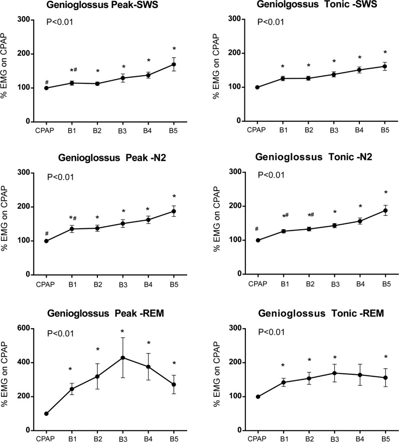
Methods: Pcrit and/or muscle data were obtained from 72 adults aged 20-64 y with and without OSA.Pcrit was determined via transient reductions in continuous positive airway pressure (CPAP) during N2, slow wave sleep (SWS) and rapid eye movement (REM) sleep. Genioglossus and tensor palatini muscle activities were measured: (1) awake with and without CPAP, (2) during stable sleep on CPAP, and (3) in response to the CPAP reductions used to quantify Pcrit.

Results: Pcrit was 4.9 ± 1.4 cmH2O higher (more collapsible) during REM versus SWS (P = 0.012), 2.3 ± 0.6 cmH2O higher during REM versus N2 (P < 0.001), and 1.6 ± 0.7 cmH2O higher in N2 versus SWS (P = 0.048). Muscle activity decreased from wakefulness to sleep and from SWS to N2 to REM sleep for genioglossus but not for tensor palatini. Pharyngeal muscle activity increased by ∼50% by breath 5 following CPAP reductions.

Conclusions: Upper airway collapsibility measured via the Pcrit technique and genioglossus muscle activity vary with sleep stage. These findings should be taken into account when performing and interpreting "passive" Pcrit measurements.

Keywords: airway anatomy; electromyography; sleep disordered breathing; upper airway physiology.


Figures

Figure 1
Figure 1
Raw data from one individual during stage 2 sleep illustrating flow limitation and genioglossus (GG) and tensor palatini (TP) electromyographic (EMG) activity during a transient reduction in continuous positive airway pressure (CPAP). EMG is presented as a moving time average (MTA) and as a percentage of the maximum activity produced during a maximum maneuver task during wakefulness. The arrow indicates the point at which CPAP was reduced. The dashed horizontal line denotes the breaths used for critical closing pressure (Pcrit) measurement. EMG activity was analyzed during the 1 min prior to the CPAP reduction and during the five breaths after the CPAP reduction. As flow limitation occurs, genioglossus and tensor palatini EMG increase.
Figure 2
Figure 2
Critical closing pressure (Pcrit) in all participants in whom Pcrit data were acquired in at least two sleep stages for these within-subjects comparisons (n = 33). Pcrit increased from slow wave sleep (SWS) to N2 nonrapid eye movement (NREM) to rapid eye movement (REM) sleep. Open circles indicate controls, closed circles indicate OSA patients. Dashed lines indicate female participants and the solid lines indicate males. The closed squares represent means ± standard error of the mean for each group. *Significant difference, P < 0.05.
Figure 3
Figure 3
Individual examples of quantification of critical closing pressure (Pcrit) from N2, SWS and REM sleep derived from 10, eight, and five continuous positive airway pressure drops respectively. Linear regression of the relationship between peak inspiratory flow (PIF) versus mask pressure (Pmask) was used to extrapolate the pressure associated with zero flow, defined as Pcrit.
Figure 4
Figure 4
Genioglossus and tensor palatini electromyography (EMG) during wakefulness off and on continuous positive airway pressure (CPAP), and on CPAP during stable slow wave sleep (SWS), N2 nonrapid eye movement (NREM), and rapid eye movement (REM) sleep. State-dependent changes occur in genioglossus and tensor palatini EMG. Conversely, genioglossus but not tensor palatini muscle activity is sleep stage dependent. Values are mean ± standard error of the mean as % of maximum EMG from all participants who had genioglossus EMG (n = 34) and tensor palatini EMG data in each of the five conditions. P values indicate significant state differences (analysis of variance). *Significant difference from wake no CPAP; $Significant difference from wake on CPAP; #Significant difference from REM.
Figure 5
Figure 5
Peak and tonic genioglossus electromyography (EMG) on continuous positive airway pressure (CPAP) within each sleep stage and during breaths (B) 1 through 5 following transient CPAP reductions used for Pcrit quantification during N2 sleep (n = 62), slow wave sleep (SWS), (n = 40) and rapid eye movement (REM) sleep (n = 37). Peak and tonic EMG increase progressively. See Table 1 for corresponding ventilatory parameters in these individuals. P values indicate significant main effect (analysis of variance). *Significant difference from the corresponding period on CPAP; #significant difference from breath 5. Values are mean ± standard error of the mean.
Figure 6
Figure 6
Tonic tensor palatini electromyography (EMG) during continuous positive airway pressure (CPAP) and breaths (B) 1 through 5 following CPAP reductions as used for Pcrit quantification during N2 sleep (n = 58), slow wave sleep (SWS) (n = 30) and rapid eye movement (REM) sleep (n = 36). Tensor palatini EMG increases during breaths 1 through 5. P values indicate a significant main effect (analysis of variance). *Significant difference compared to the corresponding period on CPAP. Values are mean ± standard error of the mean.

References

    1. Eckert DJ, White DP, Jordan AS, Malhotra A, Wellman A. Defining phenotypic causes of obstructive sleep apnea. Identification of novel therapeutic targets. Am J Respir Crit Care Med. 2013;188:996–1004. - PMC - PubMed
    1. Owens RL, Edwards BA, Eckert DJ, et al. An integrative model of physiological traits can be used to predict obstructive sleep apnea and response to non positive airway pressure therapy. Sleep. 2015;38:961–70. - PMC - PubMed
    1. Jordan AS, Wellman A, Heinzer RC, et al. Mechanisms used to restore ventilation after partial upper airway collapse during sleep in humans. Thorax. 2007;62:861–7. - PMC - PubMed
    1. Younes M. Role of Arousals in the Pathogenesis of Obstructive Sleep Apnea. Am J Respir Crit Care Med. 2004;169:623–33. - PubMed
    1. Younes M. Role of respiratory control mechanims in the pathogenesis of obstructive sleep disorders. J Appl Physiol. 2008;105:1389–405. - PubMed

Publication types