. 2015 Nov 27:6:10025.
doi: 10.1038/ncomms10025.

Janus-faced Sestrin2 controls ROS and mTOR signalling through two separate functional domains

Affiliations

Janus-faced Sestrin2 controls ROS and mTOR signalling through two separate functional domains

Hanseong Kim et al. Nat Commun. .

Abstract

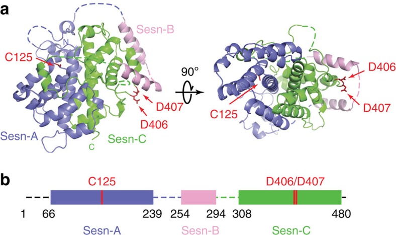
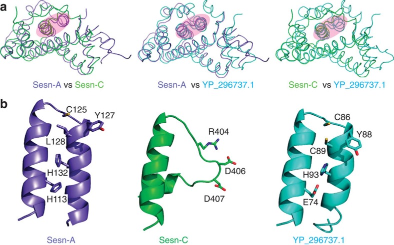
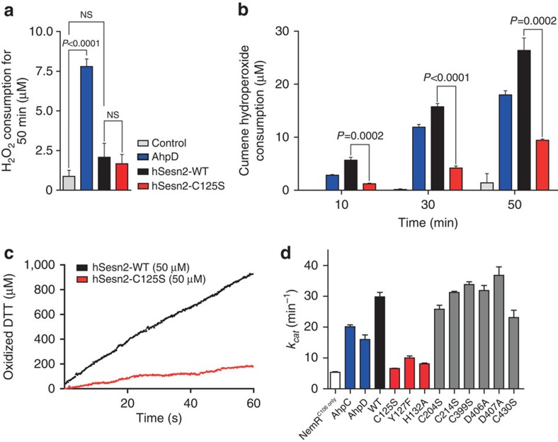
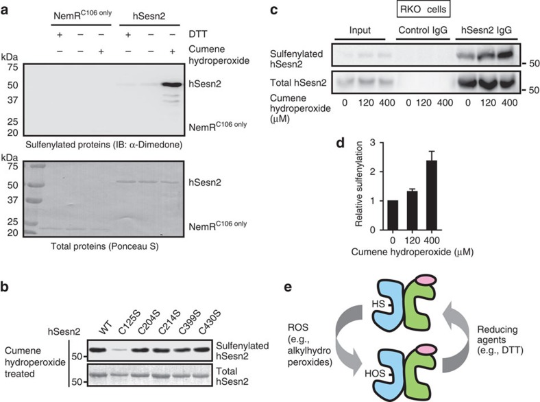
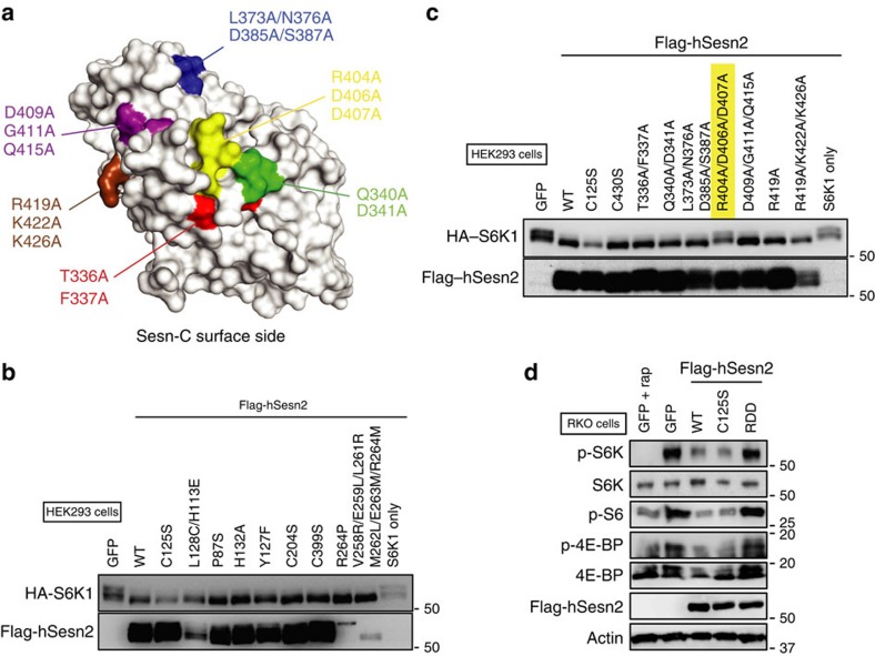
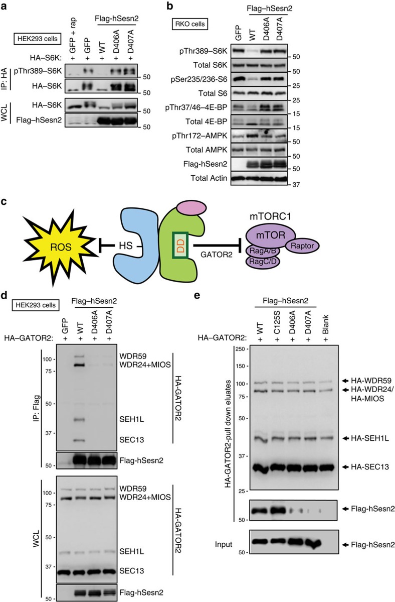
Sestrins are stress-inducible metabolic regulators with two seemingly unrelated but physiologically important functions: reduction of reactive oxygen species (ROS) and inhibition of the mechanistic target of rapamycin complex 1 (mTORC1). How Sestrins fulfil this dual role has remained elusive so far. Here we report the crystal structure of human Sestrin2 (hSesn2), and show that hSesn2 is twofold pseudo-symmetric with two globular subdomains, which are structurally similar but functionally distinct from each other. While the N-terminal domain (Sesn-A) reduces alkylhydroperoxide radicals through its helix-turn-helix oxidoreductase motif, the C-terminal domain (Sesn-C) modified this motif to accommodate physical interaction with GATOR2 and subsequent inhibition of mTORC1. These findings clarify the molecular mechanism of how Sestrins can attenuate degenerative processes such as aging and diabetes by acting as a simultaneous inhibitor of ROS accumulation and mTORC1 activation.


Figures

Figure 1
Figure 1. Crystal structure of full-length hSesn2.
(a) Ribbon diagram of full-length hSesn2. Sesn-A, Sesn-B and Sesn-C domains are in slate, pink and green, respectively. hSesn2 is composed of two globin-like α-helix-only domains (Sesn-A and Sesn-C) connected by a helix–loop–helix domain (Sesn-B) with a total of 23 helices. The overall structure is well-defined except for residues 1–65, 221–224, 240–255, 272–279, 295–307, 329–332 and 479–480. Key residues (C125, D406 and D407) in each of the globular domains are displayed in a stick model, indicated by red arrows. (b) Schematic diagram of domain organization of hSesn2. Illustrations of the protein structure used in all figures were generated with either PYMOL (Delano Scientific, LLC) or Chimera (UCSF chimera). The relative locations of C125, D406 and D407 are marked in red.
Figure 2
Figure 2. hSesn2 subdomains (Sesn-A/Sesn-C) have a structural similarity to YP_296737.1.
(a) Structural overlay between Sesn-A (slate), Sesn-C (green) and monomeric R. eutropha YP_296737.1 (cyan). The overall architectures of Sesn-A, Sesn-C and YP_296737.1 are structurally similar to each other with r.m.s. differences of 1.95 Å (Sesn-A versus Sesn-C, total 110 residues compared), 1.94 Å (Sesn-A versus YP_296737.1, 139 residues) and 2.32 Å (Sesn-C versus YP_296737.1, 104 residues). From this study, we identified two functionally active sites in each of Sesn-A and Sesn-C domains, which are highlighted in pink. (b) Structure comparison of the highlighted regions in a, which corresponds to the helix–turn–helix oxidoreductase motif of YP_296737.1. Only one cysteine is preserved in Sesn-A (Cys125), and none are found in Sesn-C.
Figure 3
Figure 3. hSesn2 is an alkylhydroperoxidase using a single catalytic cysteine in Sesn-A.
(a) hSesn2 does not show significant peroxidase activity against H2O2. Ferrous oxidation–xylenol orange (FOX) assay was used to quantify the amount of remaining H2O2 after reaction with DTT catalysed by M. tuberculosis AhpD, hSesn2-WT or hSesn2-C125S. Total H2O2 consumption amounts for the initial 50 min are measured and presented as a bar graph (n=3, mean±s.e.m.). P values were calculated using the Student's t-test. NS, non-significant (P>0.05). (b) From the FOX assay, hSesn2 shows significant peroxidase activity against cumene hydroperoxide, which is dependent on Cys125. The cumene hydroperoxide consumption for the initial 10, 30, 50 min are presented as a bar graph (n=3, mean±s.e.m.). P values were calculated using the Student's t-test. (c) Dithiothreitol (DTT)-dependent alkylhydroperoxidase activity of hSesn2 (WT and C125S mutant) towards cumene hydroperoxide was measured at 310 nm. (d) The kcat of hSesn2-WT, NemRC106 only (negative control, white), M. tuberculosis AhpC/AhpD (blue), and hSesn2-WT and hSesn2-mutants was presented as a bar graph (n=3, mean±s.e.m.).
Figure 4
Figure 4. In vitro and in vivo sulfenylation of Cys125 in hSesn2 by cumene hydroperoxides.
(a) Purified NemRC106 only and hSesn2-WT were incubated with 1 mM DTT or 120 μM cumene hydroperoxides and then treated with dimedone, which specifically labels cysteine sulfenic acids, and analysed through anti-dimedone immunoblotting (IB). Ponceau S staining was used to visualize the total levels of hSesn2 proteins. (b) Purified hSesn2 proteins of indicated Cys-to-Ser mutations were incubated with 120 μM cumene hydroperoxides. Protein sulfenylation was examined as described in a. (c) RKO cells were treated with indicated concentrations of cumene hydroperoxide for 20 min. Endogenous hSesn2 was immunopurified using hSesn2 antibodies or pre-immune immunoglobulin (IgG) and analysed by immunoblotting with a non-reducing SDS–PAGE gel. (d) Relative protein sulfenylation in c was presented as a bar graph (right panel; n=4, mean±s.e.m.). (e) Schematic diagram of the proposed reaction mechanism underlying hSesn2's peroxidase activity. Cys125 (Cys-SH) is oxidized by hydrophobic alkylhydroperoxides such as cumene hydroperoxide. The resulting sulfenic acid (Cys-SOH) is reduced directly by DTT or other unknown physiological reducing agents. Molecular weight markers are indicated in kDa.
Figure 5
Figure 5. Arg404/Asp406/Asp407 residues in Sesn-C constitute a functional site for mTORC1 regulation.
(a) The surface of Sesn-C was subdivided into five different areas (highlighted in blue, purple, yellow, green and red) that contain the most highly conserved surface residues. In addition, the residues corresponding to the formerly described putative GDI motif (Arg419, Lys422 and Lys426) are highlighted in brown. These residues were mutated into alanines as described in Supplementary Table S1. (b,c) The R404A/D406A/D407A mutation (highlighted in yellow), but none of the other mutations, abolished the mTORC1-inhibiting activity of hSesn2. mTORC1-dependent phosphorylation of S6K was monitored by electromobility retardation of HA–S6K (shifted bands). HEK293 cells were transfected with plasmid constructs expressing HA-tagged S6K1 and Flag-tagged hSesn2 of the indicated mutations. GFP or an empty vector (S6K1 only) was used as negative controls. After 48 h of transfection, cell lysates were analysed by immunoblotting of the indicated proteins. (d) RKO cells were infected with lentiviral constructs expressing GFP or Flag-tagged hSesn2 of indicated mutations. After 48 h of infection, cell lysates were analysed by immunoblotting of the indicated proteins. Rapamycin (rap, 100 nM for 24 h) was used as a positive control for mTORC1 inhibition. Molecular weight markers are indicated in kDa.
Figure 6
Figure 6. The DD motif in the Sesn-C domain of hSesn2 is responsible for direct binding to the GATOR2 complex.
(a) HEK293 cells were transfected with plasmid constructs expressing HA-tagged S6K1 and Flag-tagged hSesn2 of indicated mutations or GFP. After 48 h of transfection, HA-immunopurified protein complex (IP) and whole cell lysates (WCL) were analysed by immunoblotting of the indicated proteins. Rapamycin (rap, 100 nM for 24 h) was used as a positive control for mTORC1 inhibition. (b) RKO cells were infected with lentiviral constructs expressing GFP or Flag-tagged hSesn2 of indicated mutations. After 48 h of infection, cell lysates were analysed by immunoblotting of the indicated proteins. (c) Schematic representation of the molecular functions of hSesn2. hSesn2 is a Janus-faced molecule with two active sites in separate domains. The first site (the helix–turn–helix motif with redox-active cysteine (SH)) functions as an oxidoreductase for alkylhydroperoxide radicals, which damage critical biomolecules such as DNA. The second site (the DD motif) inhibits mTORC1 by binding to GATOR2, a recently discovered mTORC1 regulator. Inhibition of either ROS or mTORC1 can attenuate aging, and Sestrins do both. (d) Flag-tagged hSesn2 of indicated mutations was co-transfected with HA-tagged GATOR2 components (WDR59, WDR24, MIOS, SEH1L and SEC13) as indicated. Input (WCL) and Flag-immunopurified protein complex (IP) were analysed by immunoblotting. (e) WT, C125S, D406A and D407A mutants of Flag-tagged hSesn2 were purified from transiently transfected HEK293 cells. These proteins were incubated in vitro with HA–GATOR2 protein complex bound to anti-HA agarose beads. HA–GATOR2 complexes were then eluted from the beads. The pull-down eluates as well as inputs were analysed by immunoblotting of indicated proteins. Molecular weight markers are indicated in kDa.

References

    1. Lee J. H., Budanov A. V. & Karin M. Sestrins orchestrate cellular metabolism to attenuate aging. Cell Metab. 18, 792–801 (2013). - PMC - PubMed
    1. Budanov A. V., Sablina A. A., Feinstein E., Koonin E. V. & Chumakov P. M. Regeneration of peroxiredoxins by p53-regulated sestrins, homologs of bacterial AhpD. Science 304, 596–600 (2004). - PubMed
    1. Budanov A. V. & Karin M. p53 target genes sestrin1 and sestrin2 connect genotoxic stress and mTOR signalling. Cell 134, 451–460 (2008). - PMC - PubMed
    1. Lee J. H. et al. Sestrin as a feedback inhibitor of TOR that prevents age-related pathologies. Science 327, 1223–1228 (2010). - PMC - PubMed
    1. Yang Y. L. et al. SESN-1 is a positive regulator of lifespan in Caenorhabditis elegans. Exp. Gerontol. 48, 371–379 (2013). - PubMed

Publication types

MeSH terms

Associated data