. 2015 Nov 26:3:75.
doi: 10.1186/s40478-015-0254-7.

Prion-like propagation of human brain-derived alpha-synuclein in transgenic mice expressing human wild-type alpha-synuclein

Affiliations

Prion-like propagation of human brain-derived alpha-synuclein in transgenic mice expressing human wild-type alpha-synuclein

Maria E Bernis et al. Acta Neuropathol Commun. .

Abstract

Introduction: Parkinson's disease (PD) and multiple system atrophy (MSA) are neurodegenerative diseases that are characterized by the intracellular accumulation of alpha-synuclein containing aggregates. Recent increasing evidence suggests that Parkinson's disease and MSA pathology spread throughout the nervous system in a spatiotemporal fashion, possibly by prion-like propagation of alpha-synuclein positive aggregates between synaptically connected areas. Concurrently, intracerebral injection of pathological alpha-synuclein into transgenic mice overexpressing human wild-type alpha-synuclein, or human alpha-synuclein with the familial A53T mutation, or into wild-type mice causes spreading of alpha-synuclein pathology in the CNS. Considering that wild-type mice naturally also express a threonine at codon 53 of alpha-synuclein, it has remained unclear whether human wild-type alpha-synuclein alone, in the absence of endogenously expressed mouse alpha-synuclein, would support a similar propagation of alpha-synuclein pathology in vivo.

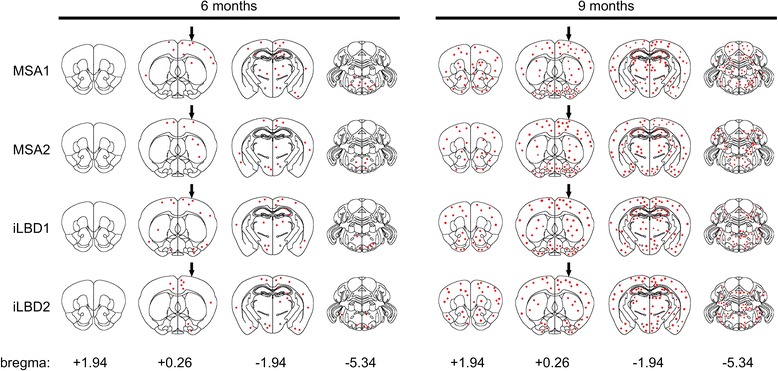
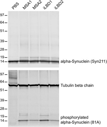
Results: Here we show that brain extracts from two patients with MSA and two patients with probable incidental Lewy body disease (iLBD) but not phosphate-buffered saline induce prion-like spreading of pathological alpha-synuclein after intrastriatal injection into mice expressing human wild-type alpha-synuclein. Mice were sacrificed at 3, 6, and 9 months post injection and analyzed neuropathologically and biochemically. Mice injected with brain extracts from patients with MSA or probable iLBD both accumulated intraneuronal inclusion bodies, which stained positive for phosphorylated alpha-synuclein and appeared predominantly within the injected brain hemisphere after 6 months. After 9 months these intraneuronal inclusion bodies had spread to the contralateral hemisphere and more rostral and caudal areas. Biochemical analysis showed that brains of mice injected with brain extracts from patients with MSA and probable iLBD contained hyperphosphorylated alpha-synuclein that also seeded aggregation of recombinant human wild-type alpha-synuclein in a Thioflavin T binding assay.

Conclusions: Our results indicate that human wild-type alpha-synuclein supports the prion-like spreading of alpha-synuclein pathology in the absence of endogenously expressed mouse alpha-synuclein in vivo.


Figures

Fig. 1
Fig. 1
Brains of MSA and probable iLBD cases contain pathogenic alpha-synuclein. Immunohistochemical staining with an antibody against phosphorylated alpha-synuclein (81A) revealed glial cytoplasmic inclusions in cortical brain tissue sections from the MSA1 patient a and in cerebellar brain tissue sections from the MSA2 patient b. Staining of cortical brain tissue sections with the same antibody also showed granular deposits of phosphorylated alpha-synuclein in the cytoplasma and in neurites of cortical neurons from the iLBD1 c and iLBD2 cases d, e. Scale bar = 50 μm
Fig. 2
Fig. 2
Biochemical analysis of alpha-synuclein in patient brains. Biochemical analysis of alpha-synuclein in patient brains shows that SDS-extractable phosphorylated alpha-synuclein was only observed in samples from patient brains a but not in a control brain b. Sequentially extracted fractions from brain homogenates of the MSA1, MSA2, iLBD1, and iLBD2 cases contain similar monomeric and oligomeric species of alpha-synuclein when probed with the EP1536Y antibody against phosphorylated alpha-synuclein (upper panel) and with the Syn211 antibody against alpha-synuclein (lower panel). Both antibodies recognize a prominent band at 64 kDa that may represent a tetramer (arrow) a and that is absent in control brain b. Sequential extractions from a control brain did not reveal phosphorylated alpha-synuclein and only showed monomeric alpha-synuclein in brain homogenate (BH), high salt (HS), and high salt-triton (HS-T) fractions (arrow) but not in the RIPA or SDS fraction b. Molecular sizes are shown in kilodalton
Fig. 3
Fig. 3
Biochemical and histological analysis of alpha-synuclein expression in wild-type and Tg(SNCA)1Nbm/J mice. Biochemical analysis with an antibody against alpha-synuclein (clone 42) that equally reacts with human as well as mouse alpha-synuclein shows that human alpha-synuclein is only very moderately overexpressed in Tg(SNCA)1Nbm/J mice in comparison to alpha-synuclein expression in wild-type mice. Equal amounts of total protein (5 μg) were loaded onto each lane of the SDS-polyacrylamide gel and show expression of alpha-synuclein in the striatum (Str), cortex (Ctx), hippocampus (Hc), brainstem (Bs), and cerebellum (Cb). Molecular sizes are shown in kilodalton a. The signal for alpha-synuclein was quantified by densitometry from western blots and normalized against tubulin and is shown as fold overexpression of alpha-synuclein in Tg(SNCA)1Nbm/J mice (n = 3) versus wild-type mice (n = 3) b. Immunohistochemistry of mouse brain sections with the same antibody (clone 42) against alpha-synuclein reveals comparable localization of alpha-synuclein in Tg(SNCA)1Nbm/J and wild-type mice c
Fig. 4
Fig. 4
Prion-like spreading of phosphorylated alpha-synuclein. The diagram depicts areas in the brains of Tg(SNCA)1Nbm/J mice with inclusion bodies that stained positive for phosphorylated alpha-synuclein at 6 and 9 months after intrastriatal injection with brain extracts from MSA or probable iLBD cases into the left striatum (arrow). Staining for phosphorylated alpha-synuclein was verified with three different antibodies, 81A, EP1536Y, and pSyn#64. Mice injected with PBS were devoid of staining for phosphorylated alpha-synuclein and inclusion bodies for up to 9 months post injection. In mice injected with brain extracts from MSA or probable iLBD cases inclusion bodies became first visible at 6 months post injection and were more abundant in the left brain hemisphere where the injection had taken place. At 9 months post injection inclusion bodies with staining for phosphorylated alpha-synuclein were more abundant and detectable to near equal amounts in both hemispheres and had also spread to more rostral and caudal areas
Fig. 5
Fig. 5
Accumulation of inclusion bodies in Tg(SNCA)1Nbm/J mice injected with brain extracts from MSA or probable iLBD cases. a Confocal images show immunofluorescence staining with the 81A antibody for phosphorylated (S129) alpha-synuclein (red) at 9 months post injection in brain sections of Tg(SNCA)1Nbm/J mice injected with brain extracts from MSA or probable iLBD cases or PBS. Neurons were stained with an antibody against tubulin beta-3 chain (green) and nuclei with DAPI (blue). Staining for phosphorylated alpha-synuclein was absent in mice injected with PBS. In contrast, mice injected with brain extracts from MSA or probable iLBD cases accumulated inclusion bodies with staining for phosphorylated alpha-synuclein in many areas of the brain, including the cortex, striatum, hippocampus, brain stem, and cerebellum. b Confocal images with orthogonal projections show that inclusion bodies with staining for phosphorylated alpha-synuclein (red) were located within neuronal cell bodies. Scale bars = 10 μm
Fig. 6
Fig. 6
Staining for phosphorylated alpha-synuclein is specific. Confocal imaging of brain sections of Tg(SNCA)1Nbm/J mice injected with brain extracts from MSA or probable iLBD cases shows that at 9 months post injection staining with the EP1536Y antibody for phosphorylated (S129) alpha-synuclein a-d and staining with the Syn211 antibody for human alpha-synuclein e-h colocalize when merged i-l. In contrast, staining with the 81A antibody for phosphorylated alpha-synuclein m-p does not colocalize with staining for neurofilament light polypeptide q-t when merged u-x. Nuclei were stained with DAPI (blue). Scale bar = 10 μm
Fig. 7
Fig. 7
Phosphorylated alpha-synuclein in inclusion bodies colocalizes with ubiquitin and sequestosome-1. Confocal imaging of brain sections of Tg(SNCA)1Nbm/J mice injected with brain extracts from MSA or probable iLBD cases shows that at 9 months post injection staining with the EP1536Y antibody for phosphorylated alpha-synuclein a-d and staining for ubiquitin e-h colocalize when merged i-l. In addition, staining with the 81A antibody for phosphorylated alpha-synuclein m-p colocalizes with staining for sequestosome-1 q-t when merged u-x. Nuclei were stained with DAPI (blue). Scale bar = 10 μm
Fig. 8
Fig. 8
Brains of mice injected with brain extracts from MSA and probable iLBD cases but not PBS accumulate phosphorylated alpha-synuclein. Brains of Tg(SNCA)1Nbm/J mice injected with brain extracts from MSA or probable iLBD cases or PBS were biochemically analyzed at 9 months post injection with antibodies against alpha-synuclein (Syn211) or phosphorylated alpha-synuclein (81A). All mouse brains contained equal amounts of sarkosyl-soluble alpha-synuclein (upper panel). Phosphorylated alpha-synuclein, however, was not detectable in mouse brains injected with PBS but in mouse brains injected with brain extracts from MSA or probable iLBD cases (lower panel). Detection of tubulin beta chain shows equal loading in all lanes. Molecular sizes are shown in kilodalton
Fig. 9
Fig. 9
Thioflavin T binding assay detects abnormal alpha-synuclein in the brains of mice injected with brain extracts from MSA and iLBD patients. Shown is the time dependent increase in Thioflavin T (ThT) fluorescence measured in relative fluorescence units (RFU) over a period of 72 h. Brain homogenates from mice injected with PBS did not seed aggregation of alpha-synuclein, suggesting that they did not contain abnormal alpha-synuclein. In contrast, brain homogenates from mice injected with brain extracts from patients with MSA1, MSA2, iLBD1, and iLBD2 lead to an increase in ThT fluorescence over time, suggesting that the brains of these mice contained abnormal alpha-synuclein that was seeding competent. Each row represents replicates from the same whole brain homogenate of mice collected at 9 months post injection

References

    1. Asi YT, Simpson JE, Heath PR, Wharton SB, Lees AJ, Revesz T, et al. Alpha-synuclein mRNA expression in oligodendrocytes in MSA. Glia. 2014;62:964–970. doi: 10.1002/glia.22653. - DOI - PMC - PubMed
    1. Ausubel FM, Brent R, Kingston RE, Moore DD, Seidman JG, Smith JA, et al. Current Protocols in Molecular Biology. New York: John Wiley & Sons; 1989.
    1. Braak H, Del Tredici K, Rub U, de Vos RA, Jansen Steur EN, Braak E. Staging of brain pathology related to sporadic Parkinson's disease. Neurobiol Aging. 2003;24:197–211. doi: 10.1016/S0197-4580(02)00065-9. - DOI - PubMed
    1. Chartier-Harlin MC, Kachergus J, Roumier C, Mouroux V, Douay X, Lincoln S, et al. Alpha-synuclein locus duplication as a cause of familial Parkinson's disease. Lancet. 2004;364:1167–1169. doi: 10.1016/S0140-6736(04)17103-1. - DOI - PubMed
    1. Collinge J, Clarke AR. A general model of prion strains and their pathogenicity. Sci. 2007;318:930–936. doi: 10.1126/science.1138718. - DOI - PubMed

Publication types

MeSH terms