

Fludarabine- (C2- methylhydroxyphosphoramide)- [anti-IGF-1R]: Synthesis and Selectively "Targeted"Anti-Neoplastic Cytotoxicity against Pulmonary Adenocarcinoma (A549)
- PMID: 26613088
- PMCID: PMC4657452
- DOI: 10.4172/2325-9604.1000129
Fludarabine- (C2- methylhydroxyphosphoramide)- [anti-IGF-1R]: Synthesis and Selectively "Targeted"Anti-Neoplastic Cytotoxicity against Pulmonary Adenocarcinoma (A549)
Abstract
Introduction: Many if not most conventional small molecular weight chemotherapeutics are highly potent against many forms of neoplastic disease. Unfortunately, majority of an administered dose unintentionally diffuses passively into normal tissues and healthy organ systems following intravenous administration. One strategy for both increasing potency and reducing dose-limited sequela is the selective "targeted" delivery of conventional chemotherapeutic agents.
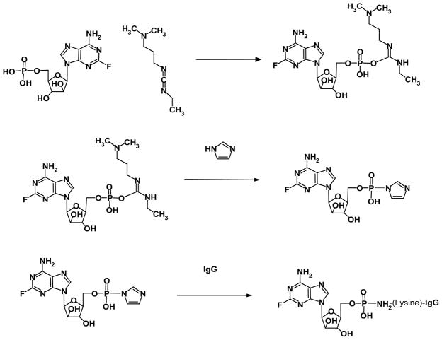
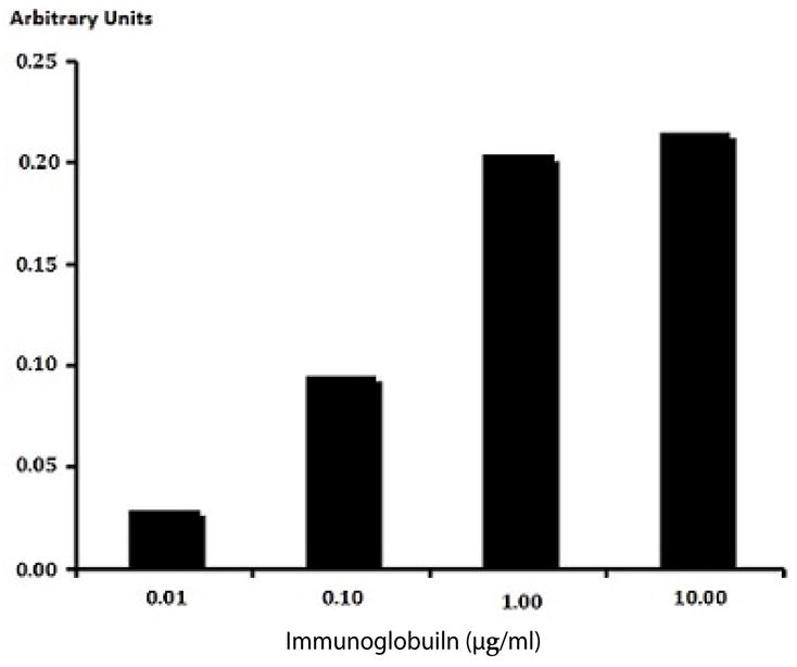
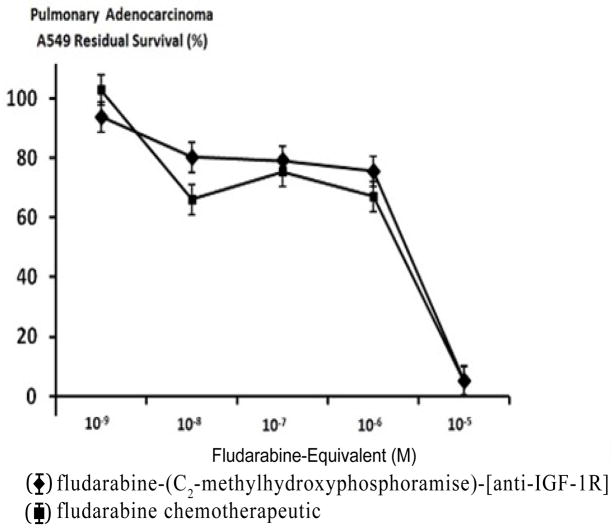
Materials and methods: The fludarabine-(C2- methylhydroxyphosphoramide)-[anti-IGF-1R] was synthesized by initially reacting fludarabine with a carbodiimide to form a fludarabine carbodiimide phosphate ester intermediate that was subsequently reacted with imidazole to create an amine-reactive fludarabine- (C2-phosphorylimidazolide) intermediate. Monoclonal anti-IGF-1R immunoglobulin was combined with the amine-reactive fludarabine- (C2-phosphorylimidazolide) intermediate resulting in the synthesis of covalent fludarabine-(C2-methylhydroxyphosphoramide)- [anti-IGF-1R] immunochemotherapeutic. Residual fludarabine and un-reacted reagents were removed by serial microfiltration (MWCO 10,000) and monitored by analytical-scale HP-TLC. Retained IGF-1R binding-avidity of fludarabine-(C2- methylhydroxyphosphoramide)-[anti-IGF-1R] was established by cell-ELISA using pulmonary adenocarcinoma cell (A549) which over-expresses IGF-1R and EGFR. Anti-neoplastic cytotoxic potency of fludarabine-(C2-methylhydroxyphosphoramide)-[anti- IGF-1R] was determined against pulmonary adenocarcinoma (A549) using an MTT-based vitality stain methodology.
Results: The fludarabine molar-incorporation-index for fludarabine- (C2-methylhydroxyphosphoramide)-[anti-IGF-R1] was 3.67:1 while non-covalently bound fludarabine was not detected by analytical scale HP-TLC following serial micro-filtration. Size-separation fludarabine-(C2-methylhydroxyphosphoramide)-[anti- IGF-1R] by SDS-PAGE with chemo luminescent autoradiography detected only a single 150-kDa band. Cell-ELISA of fludarabine- (C2-methylhydroxyphosphoramide)-[anti-IGF-1R] measuring total immunoglobulin bound to exterior surface membranes of pulmonary adenocarcinoma (A549) increased with elevations in immunoglobulin-equivalent concentrations of the covalent fludarabine immunochemotherapeutic. Between the fludarabine-equivalent concentrations of 10-10 M and 10-5 M both fludarabine-(C2- methylhydroxyphosphoramide)-[anti-IGF-1R] and fludarabine had ex-vivo anti-neoplastic cytotoxic potency levels that increased rapidly between the fludarabine-equivalent concentrations of 10-6 M and 10-5 M where cancer cell death percentages increased from 24.4% to a maximum of 94.7% respectively.
Conclusion: The molecular design and organic chemistry reaction schemes were developed for synthesizing fludarabine-(C2- methylhydroxyphosphoramide)-[anti-IGF-1R] which possessed both properties of selective "targeted" delivery and anti-neoplastic cytotoxic potency equivalent to fludarabine chemotherapeutic.
Keywords: Anti-neoplastic cytotoxicity; Cytotoxic potency; Pulmonary adenocarcinoma.
Figures


Similar articles
-
Gemcitabine-(5'-phosphoramidate)-[anti-IGF-1R]: molecular design, synthetic organic chemistry reactions, and antineoplastic cytotoxic potency in populations of pulmonary adenocarcinoma (A549).Chem Biol Drug Des. 2017 Mar;89(3):379-399. doi: 10.1111/cbdd.12845. Epub 2016 Dec 20. Chem Biol Drug Des. 2017. PMID: 27561602 Free PMC article.
-
Dexamethasone-(C21-phosphoramide)-[anti-EGFR]: molecular design, synthetic organic chemistry reactions, and antineoplastic cytotoxic potency against pulmonary adenocarcinoma (A549).Drug Des Devel Ther. 2016 Aug 12;10:2575-97. doi: 10.2147/DDDT.S102075. eCollection 2016. Drug Des Devel Ther. 2016. PMID: 27574398 Free PMC article.
-
Selectively Targeted Anti-Neoplastic Cytotoxicity of Three Immunopharmaceuticals with Covalently Bound Fludarabine, Gemcitabine and Dexamethasone Moieties Synthesized Utilizing Organic Chemistry Reactions in a Multi-Stage Regimen.Curr Pharm Des. 2018;24(11):1224-1240. doi: 10.2174/1381612823666171114155439. Curr Pharm Des. 2018. PMID: 29141539
-
Small molecule inhibitors of the IGF-1R/IR axis for the treatment of cancer.Expert Opin Investig Drugs. 2011 May;20(5):605-21. doi: 10.1517/13543784.2011.558501. Epub 2011 Mar 30. Expert Opin Investig Drugs. 2011. PMID: 21446886 Review.
-
Insulin-like growth factor signaling as a therapeutic target in pancreatic cancer.Anticancer Agents Med Chem. 2011 Jun;11(5):427-33. doi: 10.2174/187152011795677454. Anticancer Agents Med Chem. 2011. PMID: 21492074 Review.
Cited by
-
Gemcitabine-(5'-phosphoramidate)-[anti-IGF-1R]: molecular design, synthetic organic chemistry reactions, and antineoplastic cytotoxic potency in populations of pulmonary adenocarcinoma (A549).Chem Biol Drug Des. 2017 Mar;89(3):379-399. doi: 10.1111/cbdd.12845. Epub 2016 Dec 20. Chem Biol Drug Des. 2017. PMID: 27561602 Free PMC article.
-
Dexamethasone-(C21-phosphoramide)-[anti-EGFR]: molecular design, synthetic organic chemistry reactions, and antineoplastic cytotoxic potency against pulmonary adenocarcinoma (A549).Drug Des Devel Ther. 2016 Aug 12;10:2575-97. doi: 10.2147/DDDT.S102075. eCollection 2016. Drug Des Devel Ther. 2016. PMID: 27574398 Free PMC article.
References
-
- Gandhi V, Plunkett W. Cellular and clinical pharmacology of fludarabine. Clin Pharmacokinet. 2002;41:93–103. - PubMed
-
- Zinzani PL, Pellegrini C, Broccoli A, Casadei B, Argnani L, et al. Fludarabine-mitoxantrone-rituximab regimen in untreated intermediate/high-risk follicular non-Hodgkin’s lymphoma: experience on 142 patients. Am J Hematol. 2013;88:E273–276. - PubMed
Grants and funding
LinkOut - more resources
Full Text Sources
Other Literature Sources
Research Materials
Miscellaneous