Review
. 2015;9(5):239-259.
doi: 10.1159/000441988. Epub 2015 Nov 28.

The Genetic and Environmental Factors Underlying Hypospadias

Affiliations
Review

The Genetic and Environmental Factors Underlying Hypospadias

Aurore Bouty et al. Sex Dev. 2015.

Abstract

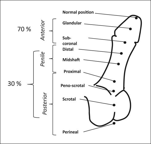
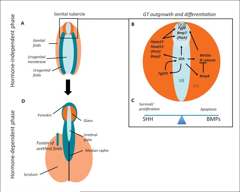
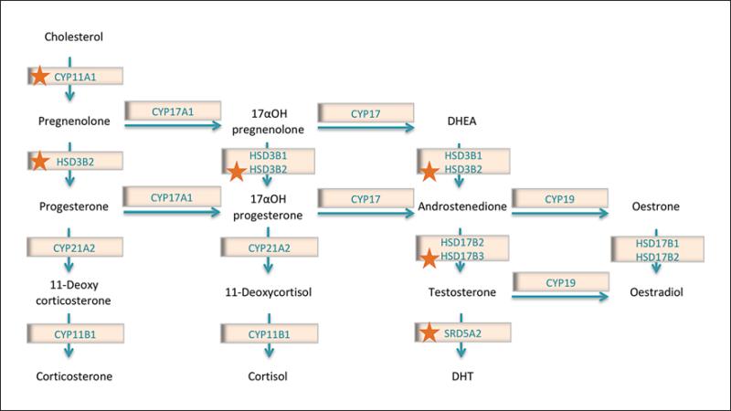
Hypospadias results from a failure of urethral closure in the male phallus and affects 1 in 200-300 boys. It is thought to be due to a combination of genetic and environmental factors. The development of the penis progresses in 2 stages: an initial hormone-independent phase and a secondary hormone-dependent phase. Here, we review the molecular pathways that contribute to each of these stages, drawing on studies from both human and mouse models. Hypospadias can occur when normal development of the phallus is disrupted, and we provide evidence that mutations in genes underlying this developmental process are causative. Finally, we discuss the environmental factors that may contribute to hypospadias and their potential immediate and transgenerational epigenetic impacts.


Figures

Fig. 1
Fig. 1
Clinical classification of the severity of hypospadias. Hypospadias severity is classified according to the position of the urethral meatus. Anterior hypospadias is also referred to as distal or minor, and posterior as proximal or severe. The respective prevalence for each form is noted.
Fig. 2
Fig. 2
The molecular pathways underlying the development of the male external genitalia. A–C The hormone-independent phase where the urogenital sinus is indifferent. This phase occurs following the division of the cloacal membrane into the urogenital sinus and anal membrane. A At this stage the tissue is made up of the urethral folds (dark blue), the urethral epithelium (light blue), the labioscrotal swellings (light orange), and the mesenchyme of the GT (dark orange). B The GT (enlarged in box) consists of a urethral plate epithelium (UE), including the distal urethral epithelium (DUE), surrounded by a bilateral mesenchyme (ME). Numerous signalling molecules contribute to GT outgrowth and differentiation during this first phase. A central cue is SHH, which is expressed in the UE, and activates its pathway via its receptor Patched (PTCH). In the bilateral mesenchyme the HH pathway activates the expression of Hoxa13 and Hoxd13, Wnt5a and its pathway (including β-catenin). SHH signalling also activates Fgf8 and Bmp7 in the DUE. SHH has a positive feedback loop with FGF10 in the mesenchyme. SHH signalling is required for both GT outgrowth and cell survival. SHH targets Fgf8 and Wnt5a are also necessary for outgrowth. Opposing this, BMP4 (and other BMPs) repress outgrowth, perhaps via their repression of Wnt5a and Fgf8. C BMP4 also represses proliferation while activating cell apoptosis, a balance that is counteracted by SHH signaling. D The later hormone-dependent stage of development after masculinisation has occurred. During masculinisation, testosterone secreted by the testis causes the GT to elongate and urogenital membrane to transform into urethral groove. Following this, all components of the penis start to close ventrally, giving rise to the median raphe. A balance between testosterone and oestrogen signalling regulates these processes. The colour of each tissue represents its origin in the previous stages.
Fig. 3
Fig. 3
The steroidogenic pathway. The synthesis of androgens occurs in the Leydig cells of the testis via the steroidogenic pathway. This pathway converts cholesterol into sex hormones, especially testosterone and dihydrotestosterone (DHT). These are necessary for the hormone-dependent stage of penis development, and disruptions in their production can cause hypospadias. Enzymes marked with a star have been implicated in human hypospadias.

References

    1. Acampora D, D'Esposito M, Faiella A, Pannese M, Migliaccio E, et al. The human HOX gene family. Nucleic Acids Res. 1989;17:10385–10402. - PMC - PubMed
    1. Achermann JC, Domenice S, Bachega TASS, Nishi MY, Mendonca BB. Disorders of sex development: effect of molecular diagnostics. Nat Rev Endocrinol. 2015;11:478–488. - PubMed
    1. Adamovic T, Chen Y, Thai HTT, Zhang X, Markljung E, et al. The p.G146A and p.P125P polymorphisms in the steroidogenic factor-1 (SF-1) gene do not affect the risk for hypospadias in Caucasians. Sex Dev. 2012;6:292–297. - PubMed
    1. Adams SV, Hastert TA, Huang Y, Starr JR. No association between maternal pre-pregnancy obesity and risk of hypospadias or cryptorchidism in male newborns. Birth Defects Res A Clin Mol Teratol. 2011;91:241–248. - PMC - PubMed
    1. Advani RJ, Yang B, Prekeris R, Lee KC, Klumperman J, Scheller RH. VAMP-7 mediates vesicular transport from endosomes to lysosomes. J Cell Biol. 1999;146:765–776. - PMC - PubMed

MeSH terms