. 2015 Nov 27;6(2):287-98.
doi: 10.1534/g3.115.024877.

SAGA DUB-Ubp8 Deubiquitylates Centromeric Histone Variant Cse4

Affiliations

SAGA DUB-Ubp8 Deubiquitylates Centromeric Histone Variant Cse4

Claudia Canzonetta et al. G3 (Bethesda). .

Abstract

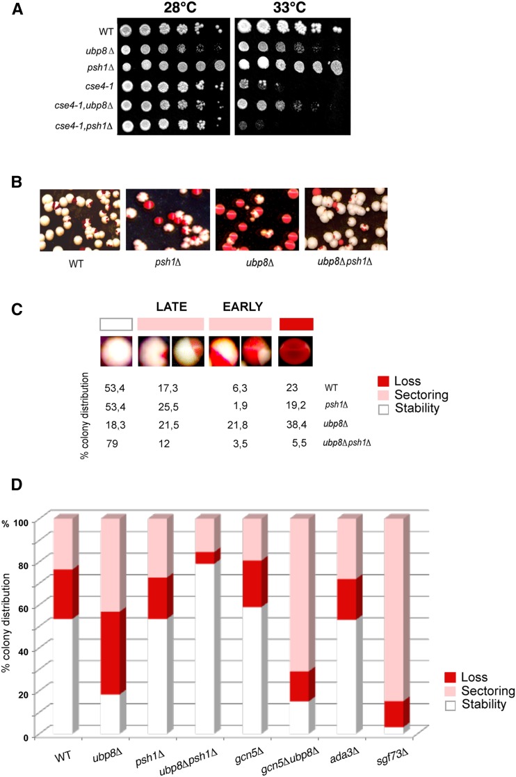
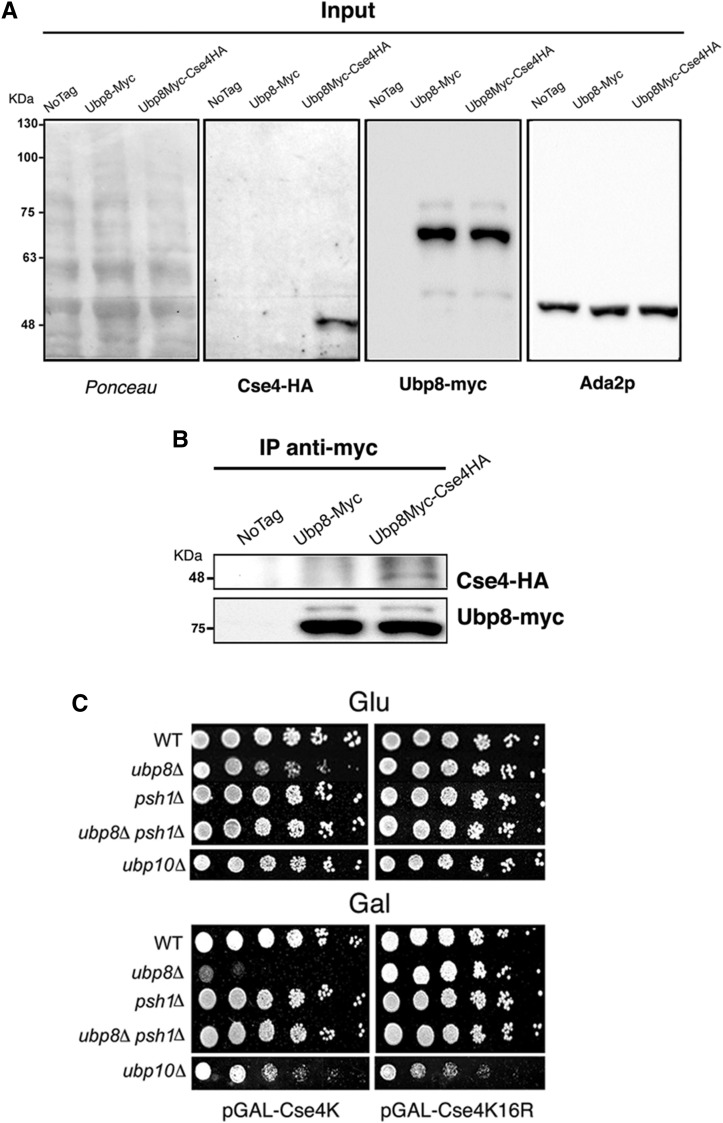
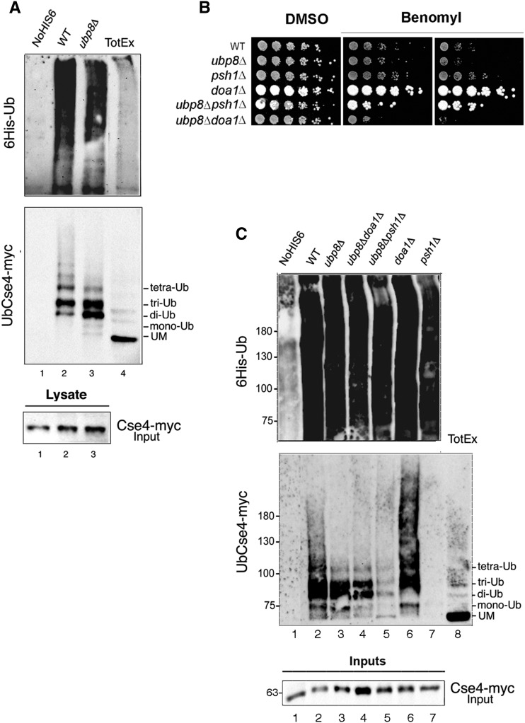
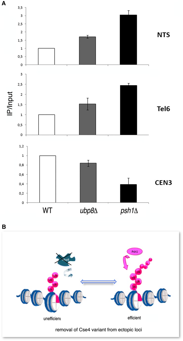
Aneuploidy, the unbalanced segregation of chromosomes during cell division, is recurrent in many tumors and the cause of birth defects and genetic diseases. Centromeric chromatin represents the chromosome attachment site to the mitotic spindle, marked by specialized nucleosomes containing a specific histone variant, CEN-H3/Cse4, in yeast. Mislocalization of Cse4 outside the centromere is deleterious and may cause aberrant chromosome behavior and mitotic loss. For this reason, ubiquitylation by the E3-ubiquitin ligase Psh1 and subsequent proteolysis tightly regulates its restricted localization. Among multiproteic machineries, the SAGA complex is not merely engaged in acetylation but also directly involved in deubiquitylation. In this study, we investigated the role of SAGA-DUB's Ubp8-driven deubiquitylation of the centromeric histone variant Cse4 in budding yeast. We found that Ubp8 works in concert with the E3-ubiquitin ligase Psh1, and that its loss causes defective deubiquitylation and the accumulation of a short ubiquitin oligomer on Cse4. We also show that lack of Ubp8 and defective deubiquitylation increase mitotic instability, cause faster Cse4 proteolysis and induce mislocalization of the centromeric histone outside the centromere. Our data provide evidence for a fundamental role of DUB-Ubp8 in deubiquitylation and the stability of the centromeric histone in budding yeast.

Keywords: DUB-Ubp8; SAGA complex; centromere; deubiquitylation; histone variant Cse4; mitotic stability.


Figures

Figure 1
Figure 1
Ubp8 interacts with CEN-H3 TS-allele cse4-1 at the genetic level and its loss increases mitotic stability. (A) Comparative growth spot assay of fivefold serial dilutions of WT, ubp8Δ; psh1Δ; cse4-1; cse4-1, ubp8Δ; cse4-1, psh1Δ strains plated on YPD and grown at permissive (28°) and nonpermissive (33°) temperatures for 2 days. Strain with TS-allele cse4-1 was assayed in the absence of DUB-Ubp8 (cse4-1, ubp8Δ) and E3-Ub ligase Psh1 (cse4-1, psh1Δ). (B) WT, ubp8Δ, psh1Δ and ubp8Δpsh1Δ strains carrying the stable ADE/LEU pRS425-1225 CEN/ARS plasmid were grown in selective medium (leu), plated on YPD, grown, and stored at 4° for red color accumulation. (C) Cell percentage (%) showing stable, sectorialized (late and early minichromosome loss), and unstable mitotic phenotype in WT, ubp8Δ, psh1Δ and ubp8Δpsh1Δ. (D) Plasmid loss rates of WT, psh1Δ, ubp8Δ, ubp8Δpsh1Δ, gcn5Δ, gcn5Δubp8Δ, sgf73Δ and ada3Δ strains. Plasmid loss is expressed as the relative percentage of white, red, and sectorialized colonies grown on plates (400-600 colonies counted for each strain). WT, wild-type; YPD, yeast extract peptone dextrose.
Figure 2
Figure 2
Ubp8 interacts with Cse4 at a physical level and acts on Cse4 lysines. (A) Inputs from WT (wild-type), Ubp8-myc, and Ubp8myc-Cse4HA strains were run on 10% PAGE, transferred on nitrocellulose membrane, stained with red-ponceau, and consecutively probed with anti-HA (Cse4-HA), anti-myc (Ubp8-myc), and anti-ADA2 (internal protein control) antibodies. (B) Coimmunoprecipitation (coIP) carried out on whole cell extracts shown in (A) indicates that Ubp8-myc is able to coIP Cse4-HA in the double-tagged strain. (C) Growth spot assay. Fivefold serial dilutions of WT, ubp8Δ, psh1Δ, ubp8Δ-psh1Δ, and ubp10Δ strains expressing pGAL-Cse4 or pGAL-Cse4K16R were plated in glucose (GLU) or inducing galactose (GAL) SC-media and grown for 5 d at 28°.
Figure 3
Figure 3
Lack of Ubp8 causes faster proteolysis of Cse4-myc rescued by concomitant deletion of the E3-Ub ligase Psh1. (A) Proteins were extracted after adding cycloheximide (CHX) (10 μg/ml) from WT (wild-type), ubp8Δ, psh1Δ, and ubp8Δpsh1Δ strains containing an integrated copy of Cse4-myc. Lysates prepared at indicated time points (min) were probed with anti-myc and anti-ADA2 antibodies. The relative amount of Cse4-myc at each time point was quantified and normalized to Ada2 levels as indicated under each lane. ADA2-normalized Cse4 protein levels at each time point of CHX treatment are presented as a fraction of the remaining amount relative to the sample t = 0. (B) The graph shows the percentage of Cse4-myc signal normalized to Ada2 in each strain at the indicated time points. WT closed diamonds, ubp8Δ closed circles, psh1Δ open squares, ubp8Δ-psh1Δ open circles. Standard errors of the means are shown. Asterisks are as follows: ** P < 0.01, * < 0.05.
Figure 4
Figure 4
SAGA-DUB Ubp8 removes a short ubiquitin oligomer from the centromeric histone Cse4. (A) Cse4-myc tagged strains containing no vector, lane 1, or a PCUP1:6HIS-Ubiquitin plasmid (WT, 2; ubp8Δ, 3). 6HIS-Ub conjugates purified on a Ni+ column were run in parallel with a wild-type (WT) total extract, lane 4. Western blot was sequentially hybridized with anti-myc and anti-6His antibodies; 1/20th of total extracts were run separately and hybridized with anti-myc for Input quantitation (lanes 1–3). (B) Growth spot assay. Fivefold serial dilutions of WT, ubp8Δ, psh1Δ, doa1Δ, and ubp8Δdoa1Δ strains were plated on YPD-DMSO and YPD-benomyl (12 and 18 μg/μl, respectively) and grown for 2 d at 28°. (C) The same procedure as in (A) was carried out with, respectively, WT (2); ubp8Δ (3); ubp8Δdoa1Δ (4); ubp8Δpsh1Δ (5); doa1Δ (6); and psh1Δ (7). Western blot was sequentially hybridized with anti-myc and anti-6His antibodies; 1/20th of total extracts were run separately and hybridized with anti-myc for Input quantitation. YPD, yeast extract peptone dextrose; DMSO, dimethyl sulfoxide.
Figure 5
Figure 5
Loss of Ubp8 increases mislocalization of Cse4 at rDNA-NTS and Tel6 regions. (A) ChIP, followed by qPCR, was performed to analyze Cse4-myc localization in wild-type (WT), ubp8Δ, and psh1Δ strains in order to determine Cse4 localization at the centromere (Cen3) and at the ectopic sites rDNA-NTS and telomere Tel6. The values on the y-axis are arbitrary units and represent the fold difference between the amount of target sequence in the immunoprecipitated fraction and the amount of target sequence in the input DNA (IP/Input). Standard error of the mean is shown. (B) Model depicting inefficient removal of Ub-oligomer-Cse4 from ectopic loci in the absence of DUB-Ubp8.

References

    1. Au W. C., Crisp M. J., DeLuca S. Z., Rando O. J., Basrai M. A., 2008. Altered dosage and mislocalization of histone H3 and Cse4p lead to chromosome loss in Saccharomyces cerevisiae. Genetics 179(1): 263–275. - PMC - PubMed
    1. Au W. C., Dawson A. R., Rawson D. W., Taylor S. B., Baker R. E., et al. , 2013. A novel role of the N terminus of budding yeast histone H3 variant Cse4 in ubiquitin-mediated proteolysis. Genetics 194(2): 513–518. - PMC - PubMed
    1. Bernad R., Sánchez P., Losada A., 2009. Epigenetic specification of centromeres by CENP-A. Exp. Cell Res. 315(19): 3233–3241. - PubMed
    1. Braun S., Madhani H. D., 2012. Shaping the landscape: mechanistic consequences of ubiquitin modification of chromatin. EMBO Rep. 13(7): 619–630. - PMC - PubMed
    1. Buvelot S., Tatsutani S. Y., Vermaak D., Biggins S., 2003. The budding yeast Ipl1/Aurora protein kinase regulates mitotic spindle disassembly. The Journal of cell biology 160(3): 329–339. - PMC - PubMed

Publication types

LinkOut - more resources