Breast MRI, digital mammography and breast tomosynthesis: comparison of three methods for early detection of breast cancer
- PMID: 26614855
- PMCID: PMC4690445
- DOI: 10.17305/bjbms.2015.616
Breast MRI, digital mammography and breast tomosynthesis: comparison of three methods for early detection of breast cancer
Abstract
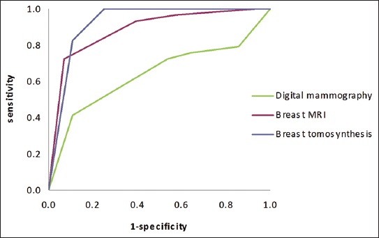
Breast cancer is the most common malignancy in women and early detection is important for its successful treatment. The aim of this study was to investigate the sensitivity and specificity of three methods for early detection of breast cancer: breast magnetic resonance imaging (MRI), digital mammography, and breast tomosynthesis in comparison to histopathology, as well as to investigate the intraindividual variability between these modalities. We included 57 breast lesions, each detected by three diagnostic modalities: digital mammography, breast MRI, and breast tomosynthesis, and subsequently confirmed by histopathology. Breast Imaging-Reporting and Data System (BI-RADS) was used for characterizing the lesions. One experienced radiologist interpreted all three diagnostic modalities. Twenty-nine of the breast lesions were malignant while 28 were benign. The sensitivity for digital mammography, breast MRI, and breast tomosynthesis, was 72.4%, 93.1%, and 100%, respectively; while the specificity was 46.4%, 60.7%, and 75%, respectively. Receiver operating characteristics (ROC) curve analysis showed an overall diagnostic advantage of breast tomosynthesis over both breast MRI and digital mammography. The difference in performance between breast tomosynthesis and digital mammography was significant (p <0.001), while the difference between breast tomosynthesis and breast MRI was not significant (p=0.20).
Figures
References
-
- American College of Radiology. ACR practice parameter for the performance of screening and diagnostic mammography Amended. 2014. [Accessed December 9 2014]. http://www.acr.org/Quality-Safety/Resources/Breast-Imaging-Resources .
-
- Chan HP, Wei J, Sahiner B, Rafferty EA, Wu T, Roubidoux M, et al. Computer-aided detection system for breast masses on digital tomosynthesis mammograms: preliminary experience. Radiology. 2005;237(3):1075–1080. http://dx.doi.org/10.1148/radiol.2373041657 . - PMC - PubMed
-
- Andersson I, Ikeda D, Zackrisson S, et al. Breast tomosynthesis and digital mammography: a comparison of breast cancer visibility and BIRADS classification in a population of cancer with subtle mammographic findings. Eur Radiol. 2008;18:2817–2825. http://dx.doi.org/10.1007/s00330-008-1076-9 . - PubMed
-
- Teertstra H, Loo C, van den Bosch M, et al. Breast tomosynthesis in clinical practice: initial results. Eur Radiol. 2010;20(1):16–24. http://dx.doi.org/10.1007/s00330-009-1523-2 . - PubMed
-
- Kontos D, Bakic PR, Carton AK, Troxel AB, Conant EF, Maidment A. Parenchymal texture analysis in digital breast tomosynthesis for breast cancer risk estimation: a preliminary study. Acad Radiol. 2009;16(3):283–298. http://dx.doi.org/10.1016/j.acra.2008.08.014 . - PMC - PubMed
Publication types
MeSH terms
LinkOut - more resources
Full Text Sources
Other Literature Sources
Medical