

Botulinum and Tetanus Neurotoxin-Induced Blockade of Synaptic Transmission in Networked Cultures of Human and Rodent Neurons
- PMID: 26615023
- PMCID: PMC4751230
- DOI: 10.1093/toxsci/kfv254
Botulinum and Tetanus Neurotoxin-Induced Blockade of Synaptic Transmission in Networked Cultures of Human and Rodent Neurons
Abstract
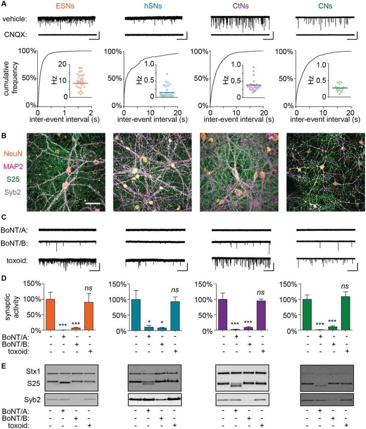
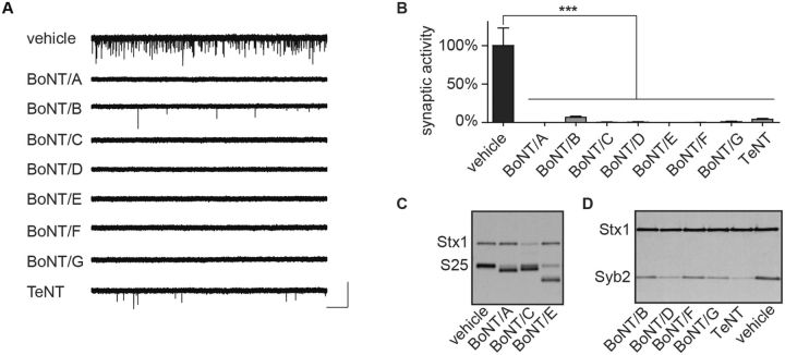
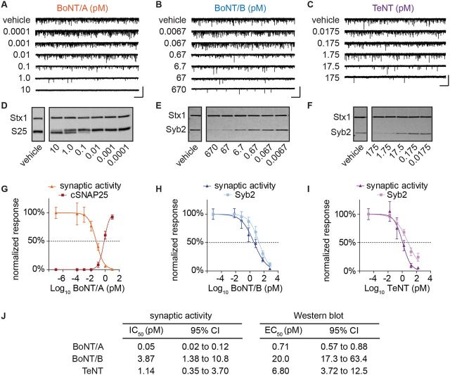
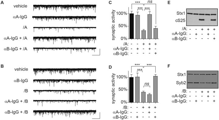
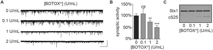
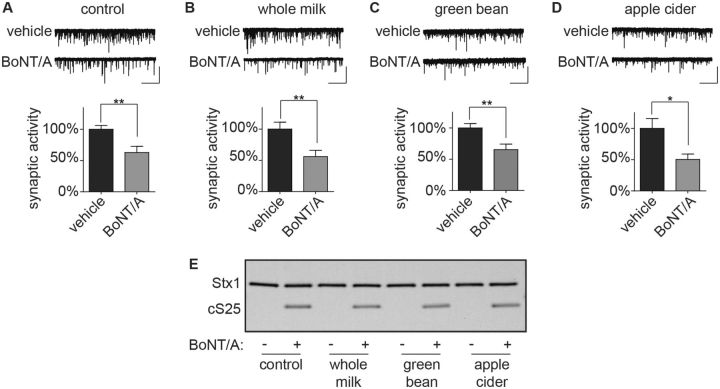
Clinical manifestations of tetanus and botulism result from an intricate series of interactions between clostridial neurotoxins (CNTs) and nerve terminal proteins that ultimately cause proteolytic cleavage of SNARE (soluble N-ethylmaleimide-sensitive factor attachment protein receptor) proteins and functional blockade of neurotransmitter release. Although detection of cleaved SNARE proteins is routinely used as a molecular readout of CNT intoxication in cultured cells, impaired synaptic function is the pathophysiological basis of clinical disease. Work in our laboratory has suggested that the blockade of synaptic neurotransmission in networked neuron cultures offers a phenotypic readout of CNT intoxication that more closely replicates the functional endpoint of clinical disease. Here, we explore the value of measuring spontaneous neurotransmission frequencies as novel and functionally relevant readouts of CNT intoxication. The generalizability of this approach was confirmed in primary neuron cultures as well as human and mouse stem cell-derived neurons exposed to botulinum neurotoxin serotypes A-G and tetanus neurotoxin. The sensitivity and specificity of synaptic activity as a reporter of intoxication was evaluated in assays representing the principal clinical and research purposes of in vivo studies. Our findings confirm that synaptic activity offers a novel and functionally relevant readout for the in vitro characterizations of CNTs. They further suggest that the analysis of synaptic activity in neuronal cell cultures can serve as a surrogate for neuromuscular paralysis in the mouse lethal assay, and therefore is expected to significantly reduce the need for terminal animal use in toxin studies and facilitate identification of candidate therapeutics in cell-based screening assays.
Keywords: botulinum toxins; electrophysiology; neurons; spontaneous postsynaptic currents; synaptic transmission; tetanus toxin.
Published by Oxford University Press on behalf of the Society of Toxicology 2015. This work is written by US Government employees and is in the public domain in the US.
Figures
Similar articles
-
Exchanging the minimal cell binding fragments of tetanus neurotoxin in botulinum neurotoxin A and B impacts their toxicity at the neuromuscular junction and central neurons.Toxicon. 2013 Dec 1;75:108-21. doi: 10.1016/j.toxicon.2013.06.010. Epub 2013 Jun 29. Toxicon. 2013. PMID: 23817019
-
Functional evaluation of biological neurotoxins in networked cultures of stem cell-derived central nervous system neurons.J Vis Exp. 2015 Feb 5;(96):52361. doi: 10.3791/52361. J Vis Exp. 2015. PMID: 25742030 Free PMC article.
-
Clostridial neurotoxins and substrate proteolysis in intact neurons: botulinum neurotoxin C acts on synaptosomal-associated protein of 25 kDa.J Biol Chem. 1996 Mar 29;271(13):7694-9. doi: 10.1074/jbc.271.13.7694. J Biol Chem. 1996. PMID: 8631808
-
Structure and function of tetanus and botulinum neurotoxins.Q Rev Biophys. 1995 Nov;28(4):423-72. doi: 10.1017/s0033583500003292. Q Rev Biophys. 1995. PMID: 8771234 Review.
-
Botulinum and Tetanus Neurotoxins.Annu Rev Biochem. 2019 Jun 20;88:811-837. doi: 10.1146/annurev-biochem-013118-111654. Epub 2018 Nov 2. Annu Rev Biochem. 2019. PMID: 30388027 Free PMC article. Review.
Cited by
-
Antidotal treatment of botulism in rats by continuous infusion with 3,4-diaminopyridine.Mol Med. 2022 Jun 3;28(1):61. doi: 10.1186/s10020-022-00487-4. Mol Med. 2022. PMID: 35659174 Free PMC article.
-
Use-dependent potentiation of voltage-gated calcium channels rescues neurotransmission in nerve terminals intoxicated by botulinum neurotoxin serotype A.Sci Rep. 2017 Nov 20;7(1):15862. doi: 10.1038/s41598-017-16064-3. Sci Rep. 2017. PMID: 29158500 Free PMC article.
-
Human-Relevant Sensitivity of iPSC-Derived Human Motor Neurons to BoNT/A1 and B1.Toxins (Basel). 2021 Aug 22;13(8):585. doi: 10.3390/toxins13080585. Toxins (Basel). 2021. PMID: 34437455 Free PMC article.
-
Engineering Botulinum Neurotoxin C1 as a Molecular Vehicle for Intra-Neuronal Drug Delivery.Sci Rep. 2017 Feb 21;7:42923. doi: 10.1038/srep42923. Sci Rep. 2017. PMID: 28220863 Free PMC article.
-
Stem cell-based approaches for developmental neurotoxicity testing.Front Toxicol. 2024 Aug 22;6:1402630. doi: 10.3389/ftox.2024.1402630. eCollection 2024. Front Toxicol. 2024. PMID: 39238878 Free PMC article. Review.
References
-
- Aarts M. M., Tymianski M. (2004). Molecular mechanisms underlying specificity of excitotoxic signaling in neurons. Curr. Mol. Med. 4, 137–147. - PubMed
-
- AOAC International (2001). AOAC official method 977.26 (sec. 17.7.01). Clostridium botulinum and its toxins in foods. Off. Methods Anal.
-
- Beaudoin G. M., 3rd, Lee S. H., Singh D., Yuan Y., Ng Y. G., Reichardt L. F., Arikkath J. (2012). Culturing pyramidal neurons from the early postnatal mouse hippocampus and cortex. Nat. Protoc. 7, 1741–1754. - PubMed
Publication types
MeSH terms
Substances
Grants and funding
LinkOut - more resources
Full Text Sources
Other Literature Sources
