. 2015 Nov 27;7(12):3472-83.
doi: 10.1093/gbe/evv240.

Genome-Wide Analysis of Evolutionary Markers of Human Influenza A(H1N1)pdm09 and A(H3N2) Viruses May Guide Selection of Vaccine Strain Candidates

Affiliations

Genome-Wide Analysis of Evolutionary Markers of Human Influenza A(H1N1)pdm09 and A(H3N2) Viruses May Guide Selection of Vaccine Strain Candidates

Sergei S Belanov et al. Genome Biol Evol. .

Abstract

Here we analyzed whole-genome sequences of 3,969 influenza A(H1N1)pdm09 and 4,774 A(H3N2) strains that circulated during 2009-2015 in the world. The analysis revealed changes at 481 and 533 amino acid sites in proteins of influenza A(H1N1)pdm09 and A(H3N2) strains, respectively. Many of these changes were introduced as a result of random drift. However, there were 61 and 68 changes that were present in relatively large number of A(H1N1)pdm09 and A(H3N2) strains, respectively, that circulated during relatively long time. We named these amino acid substitutions evolutionary markers, as they seemed to contain valuable information regarding the viral evolution. Interestingly, influenza A(H1N1)pdm09 and A(H3N2) viruses acquired non-overlapping sets of evolutionary markers. We next analyzed these characteristic markers in vaccine strains recommended by the World Health Organization for the past five years. Our analysis revealed that vaccine strains carried only few evolutionary markers at antigenic sites of viral hemagglutinin (HA) and neuraminidase (NA). The absence of these markers at antigenic sites could affect the recognition of HA and NA by human antibodies generated in response to vaccinations. This could, in part, explain moderate efficacy of influenza vaccines during 2009-2014. Finally, we identified influenza A(H1N1)pdm09 and A(H3N2) strains, which contain all the evolutionary markers of influenza A strains circulated in 2015, and which could be used as vaccine candidates for the 2015/2016 season. Thus, genome-wide analysis of evolutionary markers of influenza A(H1N1)pdm09 and A(H3N2) viruses may guide selection of vaccine strain candidates.

Keywords: evolutionary markers; influenza vaccine; influenza virus; virus evolution.


Figures

F<sc>ig</sc>. 1.—
Fig. 1.—
Evolutionary markers of influenza A(H1N1)pdm09 viruses. (A) Schematic representation of the process of identification of changes at amino acid level and their frequencies in the influenza A(H1N1)pdm09 viruses that circulated in the world during 2009–2015. (B) A scatter plot showing frequencies and distribution of these changes in concatenated viral protein sequence of the A(H1N1)pdm09 viruses from 2009 to 2015. (C) Frequencies of I354L and V344M substitutions in PB2 were analyzed in three-month intervals and plotted against virus collection dates. (D) Zoom into structure of PB2:m(7)GTP complex (PDB ID: 4PB6) showing position of 354 and 344 amino acids and their hydrophobic interaction. The distance between interacting residues is shown. (E) Phylogenetic tree constructed using A(H1N1)pdm09 strains that were circulating in the world during 2009–2015. Approximately 20 strains per year representing different geographies and collection times were used in the phylogenetic analysis. Strains were color-coded based on the year of virus collection. Full circles indicate strains that carry I354L and V344M substitutions in PB2.
F<sc>ig</sc>. 2.—
Fig. 2.—
Evolutionary markers of influenza A(H3N2) viruses. (A) Schematic representation of the process of identification of changes at amino acid level and their frequencies in the influenza A(H3N2) viruses that circulated in the world during 2009–2015. (B) A scatter plot showing frequencies and distribution of amino acid substitutions in concatenated viral protein sequence of the A(H3N2) viruses from 2009 to 2015. (C) Frequencies of D53N and Y94H in HA were analyzed in three-month intervals and plotted against virus collection dates. (D) Zoom into structure of HA:CR8020 antibody complex (PDB ID: 3SDY) showing position of sites for amino acid changes (D53N and Y94H) in HA and interacting residues of CR8020. The distance between interacting residues of HA and antibody is shown. (E) Phylogenetic tree constructed using A(H3N2) strains that were circulating in the world during 2009–2015. Approximately 20 strains per year representing different geographies and isolation times were selected. Strains were color-coded based on the year of circulation. Full circles indicate strains that carry D53N and Y94H in HA. Numbering of markers in HA is based on the protein sequence without the signal peptide.
F<sc>ig</sc>. 3.—
Fig. 3.—
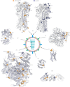
Mapping evolutionary markers of human influenza A(H1N1)pdm09 and A(H3N2) viruses on available virus protein structures. Available three-dimensional structures of individual proteins and the polymerase protein complex of influenza A viruses were used to map the positions of sites for evolutionary markers (PDB IDs: HA – 3LZG and 4FNK, NA – 1IVG, M2 – 2LY0, M1 – 3MD2, pol – 4WSB, NP – 4IRY, and NS1 – 3F5T). Sites of influenza A(H1N1)pdm09 virus are shown in orange, whereas sites of influenza A(H3N2) virus are shown in blue. Numbering starts from Met1 for all proteins except HA. Numbering of markers in HA is based on the protein sequence without the signal peptide. The schematic structure of the influenza A virion is also shown. One monomer in NA tetramer and one monomer in HA trimmer are highlighted with light-blue.
F<sc>ig</sc>. 4.—
Fig. 4.—
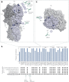
The prevalence of evolutionary markers of live A(H1N1)pdm09 viruses isolated during 2015 in A(H1N1)pdm09 vaccine strains recommended by the WHO and by the authors for 2015/2016 influenza season. (A) Frequencies of 40 selected evolutionary markers of live viruses were plotted against their positions in corresponding viral proteins. The distribution of these markers was analyzed in A(H1N1)pdm09 vaccine strains recommended by the WHO and by the authors (encircled) for 2015/2016 influenza season. Evolutionary markers are shown as black dots. Antibodies that recognize regions of viral proteins where evolutionary markers are located are also shown. (B) Mapping sites for evolutionary markers found in HA of A(H1N1)pdm09 viruses from 2015 on H1 structure (PDB ID: H1-3LZG). Mutation sites and antibodies that recognize corresponding regions on HA are shown (PDB IDs: HA:C05 – 4FP8; HA:S139/1 – 4GMS; HA:CH65 – 3SM5; HA:2D1 – 3LZG; HA:HC45 – 1QFU; HA:HB36.3 – 3R2X; HA:F-HB80.4 – 4EEF; HA:C179 – 5COR; HA:FI6V3 – 3ZTN; HA:CR6261 – 3GBN; HA:F10 – 3FKU). One monomer in HA trimmer is highlighted with light-blue. Numbering of markers in HA is based on the protein sequence without the signal peptide. (C) Mapping sites of evolutionary markers found in NA of A(H1N1)pdm09 viruses from 2015 on N1 structure (PDB ID: N1 – 4B7N). Mutation sites and antibodies that recognize corresponding regions of NA are shown (PDB IDs: NA:NC41 – 1NMB, NA:Mem5 – 2AEP). One monomer in NA tetramer is highlighted with light-blue.
F<sc>ig</sc>. 5.—
Fig. 5.—
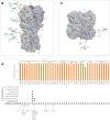
The prevalence of evolutionary markers of live A(H3N2)pdm09 viruses from 2015 in A(H3N2) vaccine strains recommended by the WHO and by the authors for 2015/2016 influenza season. (A) Frequencies of 32 selected evolutionary markers of live viruses were plotted against their positions in corresponding viral proteins. The distribution of these markers were analyzed in A(H3N2) vaccine strains recommended by the WHO and by the authors (encircled) for 2015/2016 influenza season. Evolutionary markers are shown as black dots. Antibodies that recognize regions of viral proteins where evolutionary markers are located are also shown. (B) Mapping sites for evolutionary markers found in HA of A(H3N2) viruses from 2015 on H3 structure (PDB ID: 4FNK). Mutation sites and antibodies that recognize corresponding regions on HA are shown (PDB IDs: A:F045-92 – 4O58, HA:2D1 – 3LZG, HA:S139/1 – 4GMS, HA:C05 – 4FP8, HA:FLD194 – 5A3I, HA:FI6V3 – 3ZTN). One monomer in HA trimmer is highlighted with light-blue. Numbering of markers in HA is based on the protein sequence without the signal peptide. (C) Mapping sites of evolutionary markers found in NA of A(H3N2) viruses from 2015 on N2 structure (PDB ID: N1-1IVG). Mutation sites and antibodies that recognize corresponding regions of NA are shown (PDB IDs: NA:CD6 – 4QNP, NA:Mem5 – 2AEP). One monomer in NA tetramer is highlighted with light-blue.

References

    1. Bedford T, et al. 2014. Integrating influenza antigenic dynamics with molecular evolution. eLife 3:e01914. - PMC - PubMed
    1. Bhatt S, et al. 2013. The evolutionary dynamics of influenza A virus adaptation to mammalian hosts. Philos Trans R Soc London Ser B Biol Sci. 368:20120382. - PMC - PubMed
    1. Broberg E, et al. 2015. Start of the 2014/15 influenza season in Europe: drifted influenza A(H3N2) viruses circulate as dominant subtype. Euro Surveill. 20:21023. - PubMed
    1. Corti D, et al. 2011. A neutralizing antibody selected from plasma cells that binds to group 1 and group 2 influenza A hemagglutinins. Science 333:850–856. - PubMed
    1. Drummond AJ, Ho SY, Phillips MJ, Rambaut A. 2006. Relaxed phylogenetics and dating with confidence. PLoS Biol. 4:e88. - PMC - PubMed

Publication types