TlR expression profile of human gingival margin-derived stem progenitor cells
- PMID: 26615501
- PMCID: PMC4765758
- DOI: 10.4317/medoral.20593
TlR expression profile of human gingival margin-derived stem progenitor cells
Abstract
Background: Gingival margin-derived stem/progenitor cells (G-MSCs) show remarkable periodontal regenerative potential in vivo. During regeneration, G-MSCs may interact with their inflammatory environment via toll-like-receptors (TLRs). The present study aimed to depict the G-MSCs TLRs expression profile.
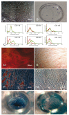
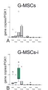
Material and methods: Cells were isolated from free gingival margins, STRO-1-immunomagnetically sorted and seeded to obtain single colony forming units (CFUs). G-MSCs were characterized for CD14, CD34, CD45, CD73, CD90, CD105, CD146 and STRO-1 expression, and for multilineage differentiation potential. Following G-MSCs' incubation in basic or inflammatory medium (IL-1β, IFN-γ, IFN-α, TNF-α) a TLR expression profile was generated.
Results: G-MSCs showed all stem/progenitor cells' characteristics. In basic medium G-MSCs expressed TLRs 1, 2, 3, 4, 5, 6, 7, and 10. The inflammatory medium significantly up-regulated TLRs 1, 2, 4, 5, 7 and 10 and diminished TLR 6 (p≤0.05, Wilcoxon-Signed-Ranks-Test).
Conclusions: The current study describes for the first time the distinctive TLRs expression profile of G-MSCs under uninflamed and inflamed conditions.
Conflict of interest statement
Figures


Similar articles
-
TLR expression profile of human alveolar bone proper-derived stem/progenitor cells and osteoblasts.J Craniomaxillofac Surg. 2017 Dec;45(12):2054-2060. doi: 10.1016/j.jcms.2017.09.007. Epub 2017 Sep 18. J Craniomaxillofac Surg. 2017. PMID: 29037921
-
Toll-like Receptor Expression Profile of Human Dental Pulp Stem/Progenitor Cells.J Endod. 2016 Mar;42(3):413-7. doi: 10.1016/j.joen.2015.11.014. Epub 2016 Jan 5. J Endod. 2016. PMID: 26769027
-
TLR-induced immunomodulatory cytokine expression by human gingival stem/progenitor cells.Cell Immunol. 2018 Apr;326:60-67. doi: 10.1016/j.cellimm.2017.01.007. Epub 2017 Jan 10. Cell Immunol. 2018. PMID: 28093098
-
Gingival Mesenchymal Stem/Progenitor Cells: A Unique Tissue Engineering Gem.Stem Cells Int. 2016;2016:7154327. doi: 10.1155/2016/7154327. Epub 2016 May 29. Stem Cells Int. 2016. PMID: 27313628 Free PMC article. Review.
-
Toll-Like Receptors and Dental Mesenchymal Stromal Cells.Front Oral Health. 2021 Apr 16;2:648901. doi: 10.3389/froh.2021.648901. eCollection 2021. Front Oral Health. 2021. PMID: 35048000 Free PMC article. Review.
Cited by
-
Response of Human Mesenchymal Stromal Cells from Periodontal Tissue to LPS Depends on the Purity but Not on the LPS Source.Mediators Inflamm. 2020 Jul 2;2020:8704896. doi: 10.1155/2020/8704896. eCollection 2020. Mediators Inflamm. 2020. PMID: 32714091 Free PMC article.
-
Gingival Mesenchymal Stem Cells: A Periodontal Regenerative Substitute.Tissue Eng Regen Med. 2025 Jan;22(1):1-21. doi: 10.1007/s13770-024-00676-8. Epub 2024 Nov 28. Tissue Eng Regen Med. 2025. PMID: 39607668 Review.
-
Effect of total sonicated Aggregatibacter actinomycetemcomitans fragments on gingival stem/progenitor cells.Med Oral Patol Oral Cir Bucal. 2018 Sep 1;23(5):e569-e578. doi: 10.4317/medoral.22661. Med Oral Patol Oral Cir Bucal. 2018. PMID: 30148477 Free PMC article.
-
Expression of Toll-like Receptors in Stem Cells of the Apical Papilla and Its Implication for Regenerative Endodontics.Cells. 2023 Oct 21;12(20):2502. doi: 10.3390/cells12202502. Cells. 2023. PMID: 37887345 Free PMC article.
-
Ascorbic Acid, Inflammatory Cytokines (IL-1β/TNF-α/IFN-γ), or Their Combination's Effect on Stemness, Proliferation, and Differentiation of Gingival Mesenchymal Stem/Progenitor Cells.Stem Cells Int. 2020 Aug 17;2020:8897138. doi: 10.1155/2020/8897138. eCollection 2020. Stem Cells Int. 2020. PMID: 32879629 Free PMC article.
References
-
- Larjava H, Wiebe C, Gallant-Behm C, Hart DA, Heino J, Häkkinen L. Exploring scarless healing of oral soft tissues. J Can Dent Assoc. 2011;77:b18. - PubMed
-
- Pitaru S, McCulloch CA, Narayanan SA. Cellular origins and differentiation control mechanisms during periodontal development and wound healing. J Periodontal Res. 1994;29:81–94. - PubMed
-
- Sempowski GD, Borrello MA, Blieden TM, Barth RK, Phipps RP. Fibroblast heterogeneity in the healing wound. Wound Repair Regen. 1995;3:120–31. - PubMed
-
- Schor SL, Ellis I, Irwin CR, Banyard J, Seneviratne K, Dolman C. Subpopulations of fetal-like gingival fibroblasts: characterisation and potential significance for wound healing and the progression of periodontal disease. Oral Dis. 1996;2:155–66. - PubMed
-
- Phipps RP, Borrello MA, Blieden TM. Fibroblast heterogeneity in the periodontium and other tissues. J Periodontal Res. 1997;32:159–65. - PubMed
Publication types
MeSH terms
Substances
LinkOut - more resources
Full Text Sources
Medical
Research Materials
Miscellaneous

