. 2016 May;49(5):276-81.
doi: 10.5483/bmbrep.2016.49.5.241.

Corosolic acid ameliorates acute inflammation through inhibition of IRAK-1 phosphorylation in macrophages

Affiliations

Corosolic acid ameliorates acute inflammation through inhibition of IRAK-1 phosphorylation in macrophages

Seung-Jae Kim et al. BMB Rep. 2016 May.

Abstract

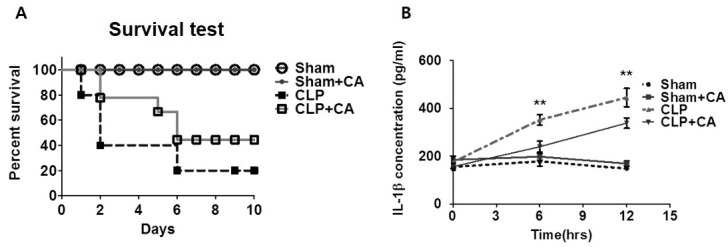
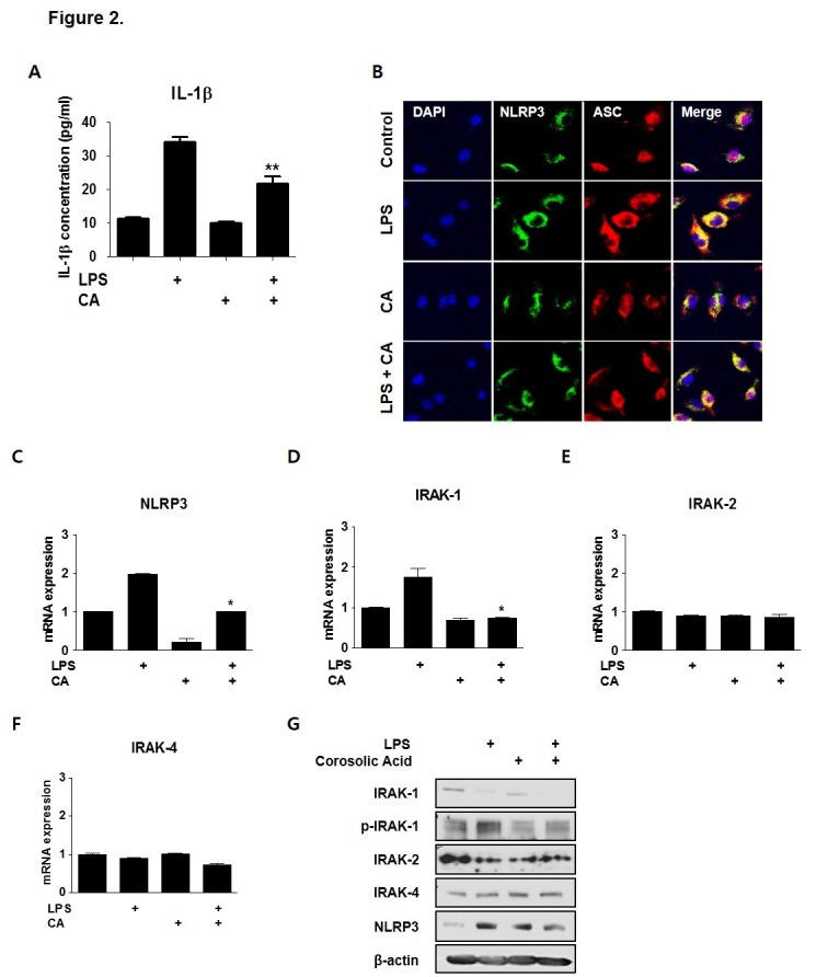
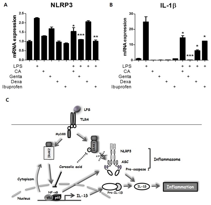
Corosolic acid (CA), a triterpenoid compound isolated from Lagerstroemia speciosa L. (Banaba) leaves, exerts anti-inflammatory effects by regulating phosphorylation of interleukin receptor-associated kinase (IRAK)-2 via the NF-κB cascade. However, the protective effect of CA against endotoxic shock has not been reported. LPS (200 ng/mL, 30 min) induced phosphorylation of IRAK-1 and treatment with CA (10 μM) significantly attenuated this effect. In addition, CA also reduced protein levels of NLRP3 and ASC which are the main components of the inflammasome in BMDMs. LPS-induced inflammasome assembly through activation of IRAK-1 was down-regulated by CA challenge. Treatment with Bay11-7082, an inhibitor of IκB-α, had no effect on CA-mediated inhibition of IRAK-1 activation, indicating that CA-mediated attenuation of IRAK-1 phosphorylation was independent of NF-κB signaling. These results demonstrate that CA ameliorates acute inflammation in mouse BMDMs and CA may be useful as a pharmacological agent to prevent acute inflammation. [BMB Reports 2016; 49(5): 276-281].


Figures

Fig. 1.
Fig. 1.. Effect of CA on mice subjected to cecal ligation and puncture (CLP) surgery and inflammasome assembly. (A) Survival of mice after CLP with or without CA (2 μg/kg). Mortality of Sham (n = 14), Sham+CA (n = 14), CLP (n = 14), and CLP+CA mice (n = 15) was monitored for 10 days after CLP surgery. (B) IL-1β secretion in vivo. Serum samples were collected 0, 6, and 12 h after surgery. The IL-1β concentration was measured by ELISA. The survival rate was estimated by the Kaplan-Meier method and compared by using the log-rank test. **P < 0.01. CLP versus CLP+CA, Sham, sham laparotomy; CLP, one puncture in the cecum; CLP+CA, one puncture in the cecum and CA injection.
Fig. 2.
Fig. 2.. CA regulates inflammasome assembly and IRAK-1 phosphorylation in BMDMs. BMDMs were isolated from mice and exposed to LPS (200 ng/mL) and/or CA (6 μM) for 30 min. (A) IL-1β secretion in vitro. BMDMs were treated with LPS and/or CA for 6 h and supernatants were collected. IL-1β secretion was measured by ELISA. (B) CA prevents inflammasome assembly. Immunocytochemistry for the inflammasome complex. BMDMs were treated with LPS and/or CA for 30 min, after which the treated cells were stained with fluorescence-labeled antibodies. Fluorescence was detected using confocal laser microscopy and LSM 3 EXCITER software. qPCR was used to measure mRNA levels of NLRP3 (C), IRAK1 (D), IRAK2 (E), and IRAK4 (F). (G) Protein samples were assessed by immunoblotting with the indicated antibodies. Graphs illustrate the mean ± SEM from 3 independent experiments. *P < 0.05, **P < 0.01 versus the relevant control group.
Fig. 3.
Fig. 3.. Relationship between CA and NF-kB signaling in acute inflammation. BMDMs were isolated from mice and exposed to LPS (200 ng/mL) and/or CA (6 μM), Bay11-7082 (10 μM), or an IRAK inhibitor (2.5 μM) for 30 min. qPCR was used to measure mRNA levels of NLRP3 (A), IRAK1 (B), IRAK2 (C), and IRAK4 (D). *P < 0.05, **P < 0.01 versus the relevant control group.
Fig. 4.
Fig. 4.. Effect of CA on LPS-mediated inflammation. (A-B) Effect of CA as an anti-inflammatory drug. BMDMs were isolated from mice and exposed to LPS (200 ng/mL) and/or gentamicin (10 μM), dexamethasone (10 μM), ibuprofen (10 μM), or CA (6 μM) for 30 min. qPCR was used to measure mRNA levels of NLRP3 and IL-1β. (C) Schematic summary of the role of CA in regulating early-phage inflammation. Graphs illustrate the mean ± SEM from 3 independent experiments. *P < 0.05, **P < 0.01, ***P < 0.001 versus the relevant control group.

References

    1. Kushner I. The phenomenon of the acute phase response. Ann N Y Acad Sci. (1982);389:39–48. doi: 10.1111/j.1749-6632.1982.tb22124.x. - DOI - PubMed
    1. Sherwood ER, Toliver-Kinsky T. Mechanisms of the inflammatory response. Best Pract Res Clin Anaesthesiol. (2004);18:385–405. doi: 10.1016/j.bpa.2003.12.002. - DOI - PubMed
    1. Kim KK, Jin SH, Lee BJ. Herpes virus entry mediator signaling in the brain is imperative in acute inflammation-induced anorexia and body weight loss. Endocrinol Metab (Seoul) (2013);28:214–220. doi: 10.3803/EnM.2013.28.3.214. - DOI - PMC - PubMed
    1. Rock KL, Kono H. The inflammatory response to cell death. Annu Rev Pathol. (2008);3:99–126. doi: 10.1146/annurev.pathmechdis.3.121806.151456. - DOI - PMC - PubMed
    1. Akira S. Toll-like receptors and innate immunity. Adv Immunol. (2001);78:1–56. - PubMed

MeSH terms