Review
. 2016 Feb;27(1):217-36.
doi: 10.1016/j.pmr.2015.08.006. Epub 2015 Oct 20.

An Evidence-Based Videotaped Running Biomechanics Analysis

Affiliations
Review

An Evidence-Based Videotaped Running Biomechanics Analysis

Richard B Souza. Phys Med Rehabil Clin N Am. 2016 Feb.

Abstract

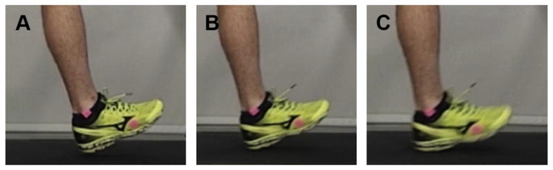
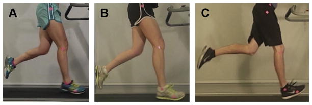
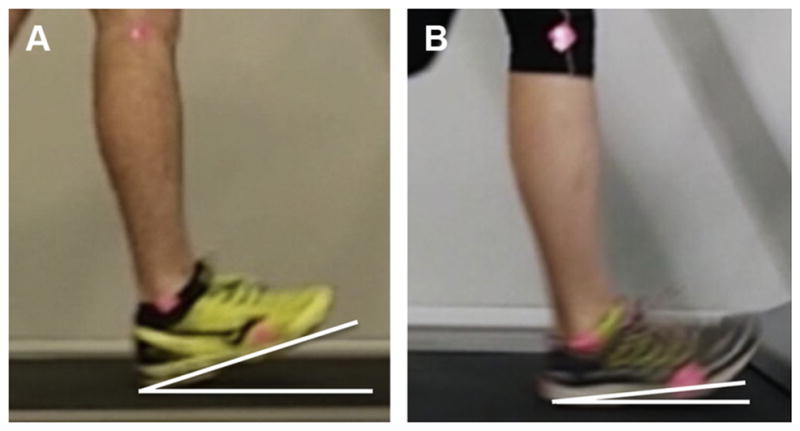
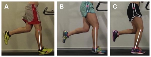
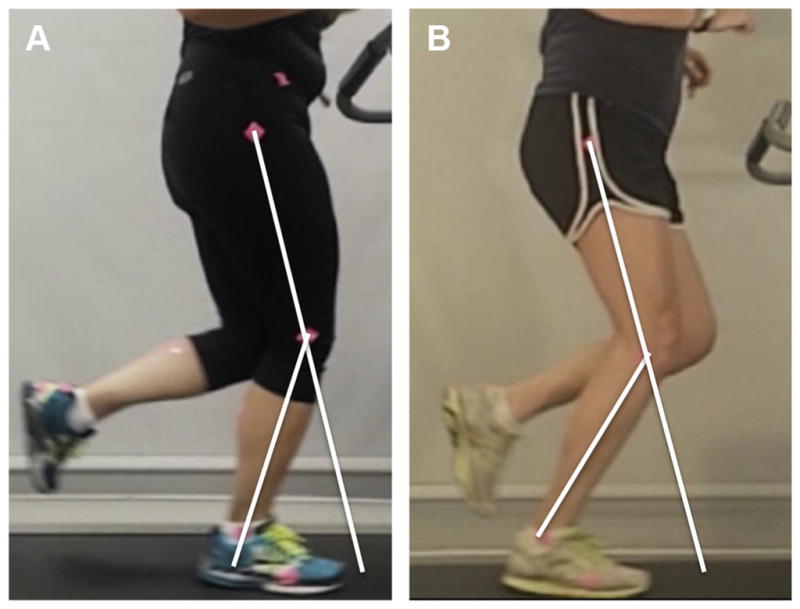
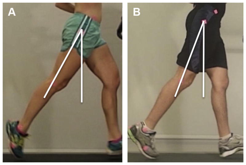
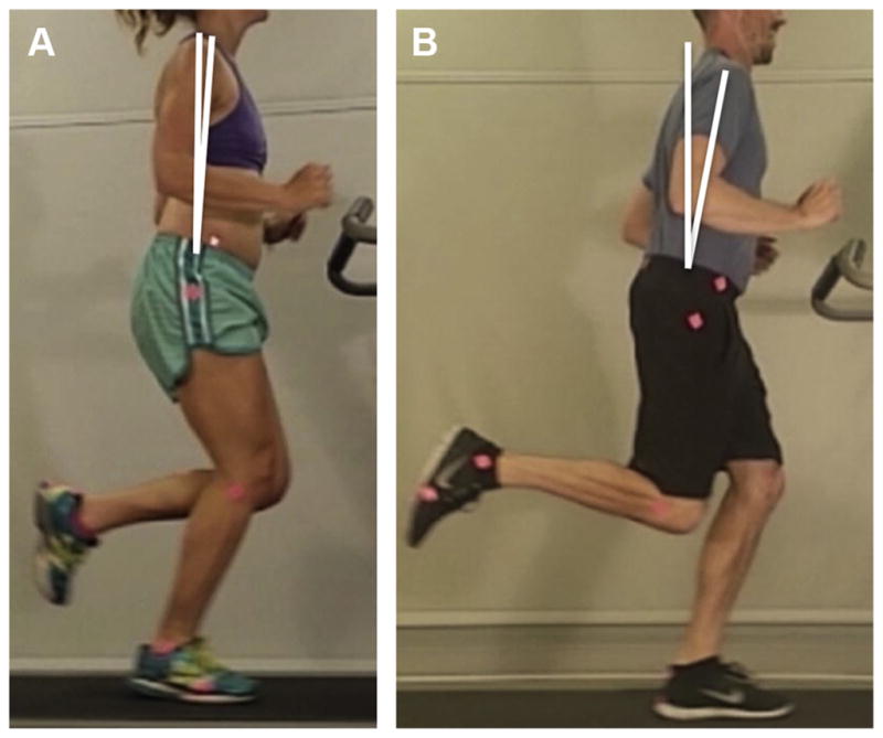
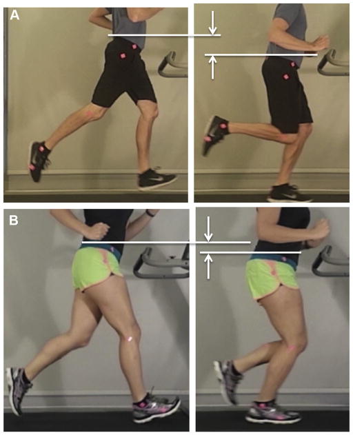
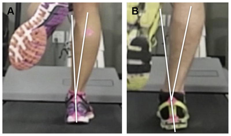
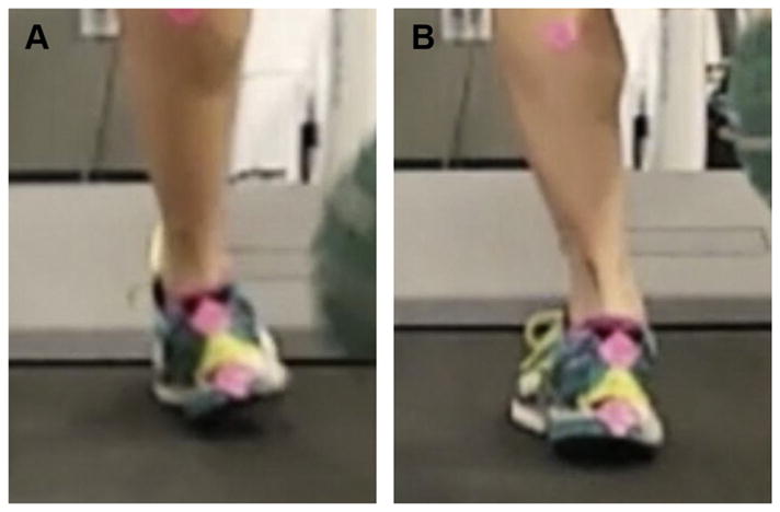
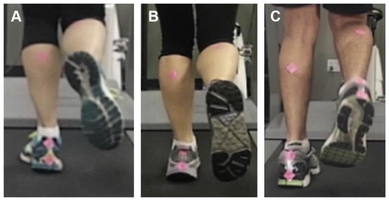
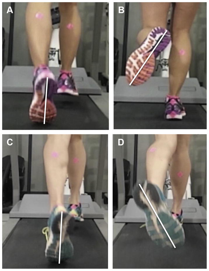
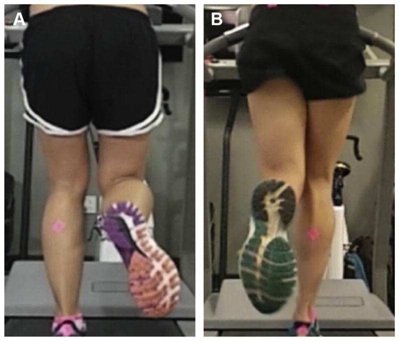
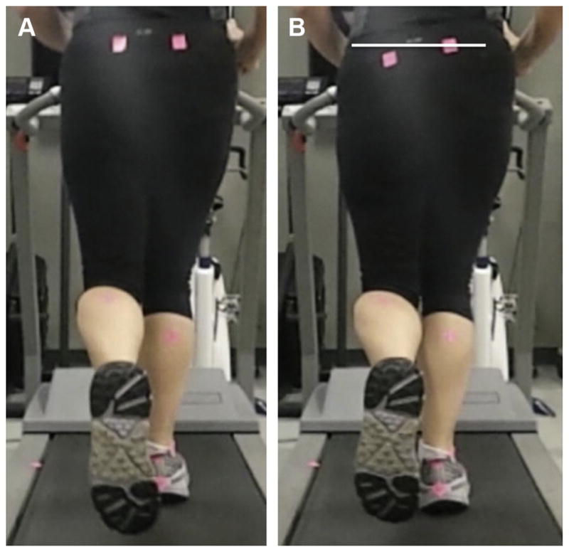
Running biomechanics play an important role in the development of injuries. Performing a running biomechanics analysis on injured runners can help to develop treatment strategies. This article provides a framework for a systematic video-based running biomechanics analysis plan based on the current evidence on running injuries, using 2-dimensional (2D) video and readily available tools. Fourteen measurements are proposed in this analysis plan from lateral and posterior video. Identifying simple 2D surrogates for 3D biomechanic variables of interest allows for widespread translation of best practices, and have the best opportunity to impact the highly prevalent problem of the injured runner.

Keywords: Biomechanics; Form; Injuries; Motion analysis; Observational; Running; Video analysis.


Figures

Fig. 1
Fig. 1
Key phases of running. (A) The end of terminal swing is identified as to the foot remains elevated from the treadmill, just before initial contact. (B) Initial contact is identified as the first frame when the foot hits the ground. (C) Loading response is identified as the first frame in which the runner’s weight is being transferred onto the lead leg and is characterized by the presence of shoe deformation.
Fig. 2
Fig. 2
Foot strike patterns. (A) Forefoot strike. (B) Midfoot strike. (C) Rear foot strike.
Fig. 3
Fig. 3
Foot inclination angle. (A) A relatively high foot inclination angle in comparison with a horizontal line. (B) A relatively low foot inclination angle.
Fig. 4
Fig. 4
Tibia angle. (A) Extended tibia. (B) Vertical tibia. (C) Flexed tibia.
Fig. 5
Fig. 5
Knee flexion during stance. (A) A runner demonstrating limited knee flexion during stance and (B) a normal amount of knee flexion during stance.
Fig. 6
Fig. 6
Hip extension during late stance. (A) Runner with normal hip extension. (B) Runner with limited hip extension.
Fig. 7
Fig. 7
Trunk lean. (A) A relatively upright trunk posture and (B) a runner a forward trunk lean.
Fig. 8
Fig. 8
Overstriding, measured at loading response. (A) A runner demonstrating normal stride mechanics and (B) a runner demonstrating an overstride, characterized by a vertical line through the lateral malleolus falling anterior to the runners pelvis.
Fig. 9
Fig. 9
Vertical displacement of the center of mass. (A) A bounding runner characterized by a large vertical displacement and (B) a relatively efficient runner with less vertical displacement.
Fig. 10
Fig. 10
Heel eversion. (A) A runner with normal alignment of the heel during running and (B) a runner with mildly excessive heel eversion during running.
Fig. 11
Fig. 11
Rate of heel eversion. A runner demonstrating excessive heel eversion and a high rate of heel eversion excursion. (A) Initial contact with the runner’s heel in an inverted position and (B) 20 milliseconds later the heel has rotated more than 20° into eversion.
Fig. 12
Fig. 12
Foot progression. (A) Normal foot progression angle. (B) Mild toe-in abnormality. (C) Severe toe-in abnormality.
Fig. 13
Fig. 13
Heel whips. Medial heel whip at initial swing (A) and maximum whip angle (B) and lateral heel whip at initial swing (C) and maximum whip (D).
Fig. 14
Fig. 14
Knee window. (A) Normal knee window and (B) “closed” knee window.
Fig. 15
Fig. 15
Excessive pelvic drop. (A) At initial contact the runner’s pelvis is fairly level and (B) during stance demonstrating excessive pelvis drop.

References

    1. Taunton JE, Ryan MB, Clement DB, et al. A retrospective case-control analysis of 2002 running injuries. Br J Sports Med. 2002;36(2):95–101. - PMC - PubMed
    1. Noehren B, Pohl MB, Sanchez Z, et al. Proximal and distal kinematics in female runners with patellofemoral pain. Clin Biomech. 2012;27(4):366–71. - PMC - PubMed
    1. Noehren B, Schmitz A, Hempel R, et al. Assessment of strength, flexibility, and running mechanics in men with iliotibial band syndrome. J Orthop Sports Phys Ther. 2014;44(3):217–22. - PMC - PubMed
    1. Milner CE, Hamill J, Davis I. Are knee mechanics during early stance related to tibial stress fracture in runners? Clin Biomech. 2007;22(6):697–703. - PubMed
    1. Edwards WB, Taylor D, Rudolphi TJ, et al. Effects of stride length and running mileage on a probabilistic stress fracture model. Med Sci Sports Exerc. 2009;41(12):2177–84. - PubMed

MeSH terms