. 2015 Nov 30:5:17375.
doi: 10.1038/srep17375.

Isolation and Identification of Post-Transcriptional Gene Silencing-Related Micro-RNAs by Functionalized Silicon Nanowire Field-effect Transistor

Affiliations

Isolation and Identification of Post-Transcriptional Gene Silencing-Related Micro-RNAs by Functionalized Silicon Nanowire Field-effect Transistor

Kuan-I Chen et al. Sci Rep. .

Abstract

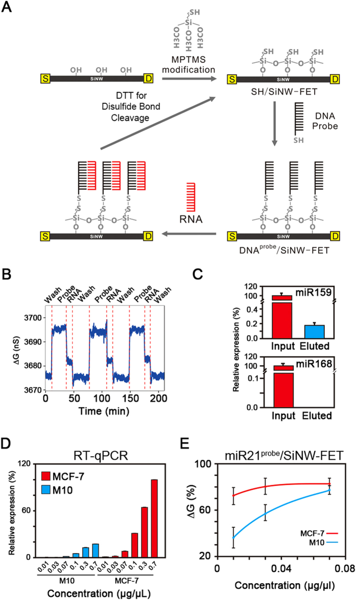
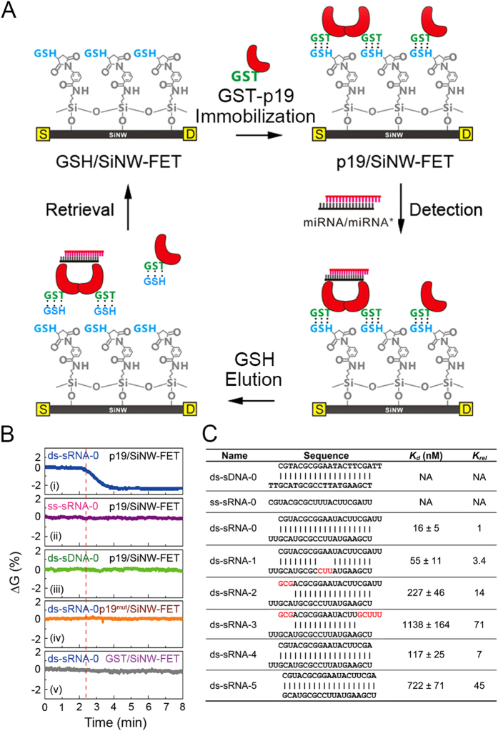
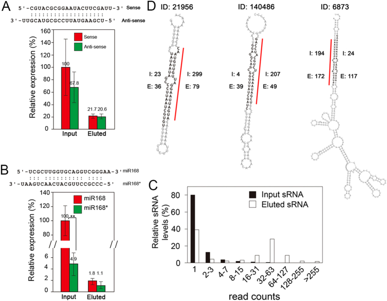
Many transcribed RNAs are non-coding RNAs, including microRNAs (miRNAs), which bind to complementary sequences on messenger RNAs to regulate the translation efficacy. Therefore, identifying the miRNAs expressed in cells/organisms aids in understanding genetic control in cells/organisms. In this report, we determined the binding of oligonucleotides to a receptor-modified silicon nanowire field-effect transistor (SiNW-FET) by monitoring the changes in conductance of the SiNW-FET. We first modified a SiNW-FET with a DNA probe to directly and selectively detect the complementary miRNA in cell lysates. This SiNW-FET device has 7-fold higher sensitivity than reverse transcription-quantitative polymerase chain reaction in detecting the corresponding miRNA. Next, we anchored viral p19 proteins, which bind the double-strand small RNAs (ds-sRNAs), on the SiNW-FET. By perfusing the device with synthesized ds-sRNAs of different pairing statuses, the dissociation constants revealed that the nucleotides at the 3'-overhangs and pairings at the terminus are important for the interactions. After perfusing the total RNA mixture extracted from Nicotiana benthamiana across the device, this device could enrich the ds-sRNAs for sequence analysis. Finally, this bionanoelectronic SiNW-FET, which is able to isolate and identify the interacting protein-RNA, adds an additional tool in genomic technology for the future study of direct biomolecular interactions.


Figures

Figure 1
Figure 1. Detection of the endogenous miRNA by SiNW-FET.
(A) A flow diagram of a reusable DNAprobe/SiNW-FET device. The MPTMS-modified SiNW-FET (SH/SiNW-FET) provides reversible disulfide bonding sites for the DNAprobe tagged with a thiol group at the 3′ end (DNAprobe/SiNW-FET). After the targeted miRNAs bind to the DNAprobe/SiNW-FET, the bound DNAprobe-miRNA complex can be eluted by flushing dithiothreitol (DTT) to break the disulfide bond, returning the device surface to SH/SiNW-FET. (B) The electrical conductance changes (ΔGs) of SH/SiNW-FET during repeated cycles of DTT-buffer washing, miR159probe modification, and RNA binding (0.3 μg/μL total RNA extracted from Arabidopsis). (C) miRNA159 in the Input and Eluted mixtures. After using miR159probe/SiNW-FET, the relative amounts of miR159 (upper panel) and miR168 (lower panel) in the bound fraction (Eluted) to the total RNA (Input) extracted from Arabidopsis were analyzed by RT-qPCR. (D,E) Comparison of the detection limits between RT-qPCR and SiNW-FET. We determined the amounts of miR21 expressed in different concentrations of total RNA extracted from cancer cell lines, MCF-7 and M10, by (D) RT-qPCR with specific primers or (E) miR21probe/SiNW-FET.
Figure 2
Figure 2. Discriminating the secondary structures of ds-sRNA by p19/SiNW-FET.
(A) A flow diagram of a reusable p19/SiNW-FET using the GSH/GST association-dissociation. The process includes the immobilization of GST-p19 on a GSH/SiNW-FET to form p19/SiNW-FET, the application of ds-sRNA (miRNA/miRNA*) to bind p19, and the elution of the GST-p19-ds-sRNA complexes with ≥1 mM GSH. (B) The responses of p19/SiNW-FET to various forms of nucleic acids. The normalized ∆Gs were measured by introducing 1 μM of a 21-nucleotide solution in (i). perfectly matched ds-sRNA form (ds-sRNA-0, the structure depicted in (C)); (ii). ss-sRNA form (ss-sRNA-0); (iii). ds-sDNA form (ds-sDNA-0) to a p19/SiNW-FET; (iv). p19 replaced by a binding-deficient mutant (p19mut); and (v). p19 absent in the tests. The vertical red-dotted line indicates the addition of samples. (C) The dissociation constant (Kd) of p19 to various 21-nucleotide nucleic acids with different mismatch pairings. The ΔGs of p19/SiNW-FET to different concentrations of nucleic acids were used to determine the Kd values (Supplementary Fig. S5), which were then normalized to that of the ds-sRNA-0 (Krel). Nucleotides marked in red indicate the mispaired bases.
Figure 3
Figure 3. p19/SiNW-FET sequestration enriches the ds-sRNAs.
We perfused the p19/SiNW-FET with sRNA (Total) from synthesized ds-sRNA-0 (A) or extracted RNA from Arabidopsis (B) or Nicotiana benthamiana (C); we then eluted the bound sRNA (Eluted) from p19/SiNW-FET and analyzed the relative amounts of the designated sRNA by (A,B) RT-qPCR or (C) a deep sequencer. (A) The relative amounts of sense and anti-sense strands of ds-sRNA-0 in the total and eluted fractions. (B) The relative amounts of miR168 (sense) and miR168* (anti-sense) in the extracted and bound fractions. (C) Proportions of the analyzed sRNAs with different read counts. The number of each sRNA sequenced was counted (read counts) and binned with a power of 2; the number of sRNAs in each binning group was normalized to the total number of sRNA counted. (D) Predicted structures of three pre-miRNAs yielding the corresponding paired ds-sRNAs captured on p19/SiNW-FET. According to the genome of N. benthamiana, nine genes (Table S1) might transcribe pre-miRNAs which have 2nd structures yielding paired ds-sRNAs (bases in bold) identified from the Elute fraction. The red lines indicate the matured miRNA forms. The digits beside each sRNA segment (bases in bold) are the counts of the corresponding sRNA segment appearing in the Input (I) or Eluted (E) sRNA sample. The data (mean ± standard deviations) were the averages of three independent experiments and ** indicates the p-value < 0.01. A magnified image of Fig. 3D is shown in Supplementary Fig. S7 for easier reading.

References

    1. Hannon G. J. RNA interference. Nature 418, 244–251 (2002). - PubMed
    1. Tomari Y. & Zamore P. D. Perspective: machines for RNAi. Gene Dev. 19, 517–529 (2005). - PubMed
    1. Bartel D. P. MicroRNAs: Genomics, biogenesis, mechanism, and function. Cell 116, 281–297 (2004). - PubMed
    1. Wu H.-W., Lin S.-S., Chen K.-C., Yeh S.-D. & Chua N.-H. Discriminating mutations of HC-Pro of zucchini yellow mosaic virus with differential effects on small RNA pathways involved in viral pathogenicity and symptom development. Mol. Plant-Microbe Interact. 23, 17–28 (2010). - PubMed
    1. Wu R.-M., Wood M., Thrush A., Walton E. F. & Varkonyi-Gasic E. Real-Time PCR quantification of plant miRNAs using Universal ProbeLibrary Technology. Biochemica 2, 12–15 (2007).

Publication types