Review
. 2015 Dec;38(12):763-775.
doi: 10.1016/j.tins.2015.10.004. Epub 2015 Nov 24.

Grid Cells and Place Cells: An Integrated View of their Navigational and Memory Function

Affiliations
Review

Grid Cells and Place Cells: An Integrated View of their Navigational and Memory Function

Honi Sanders et al. Trends Neurosci. 2015 Dec.

Abstract

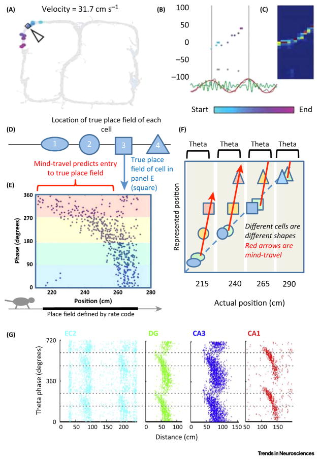
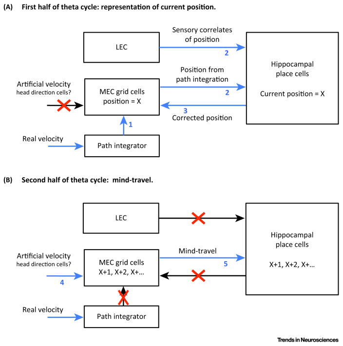
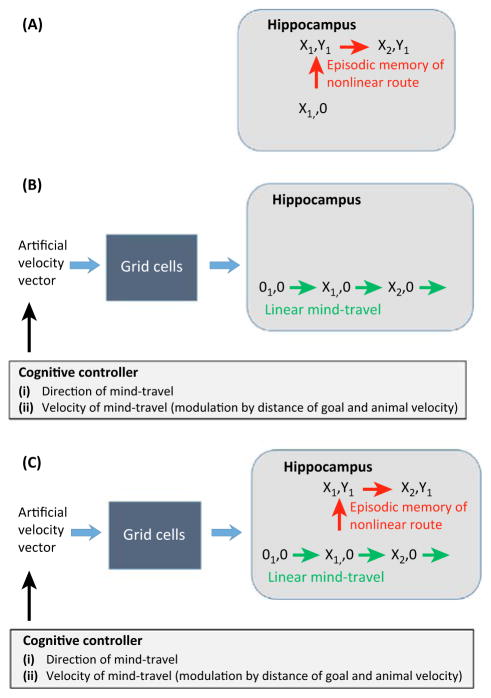
Much has been learned about the hippocampal/entorhinal system, but an overview of how its parts work in an integrated way is lacking. One question regards the function of entorhinal grid cells. We propose here that their fundamental function is to provide a coordinate system for producing mind-travel in the hippocampus, a process that accesses associations with upcoming positions. We further propose that mind-travel occurs during the second half of each theta cycle. By contrast, the first half of each theta cycle is devoted to computing current position using sensory information from the lateral entorhinal cortex (LEC) and path integration information from the medial entorhinal cortex (MEC). This model explains why MEC lesions can abolish hippocampal phase precession but not place fields.


Figures

Figure I
Figure I. MEC Grid Cell Network
Grid cells in the network are indicated by the small dots; color represents level of activation. Actual position in the environment can be constructed from combining the representations from several such networks (modules) having different spatial scales and rotation. Laterally-connecting cells (not shown) move the position of all bumps (left, before movement; right, after) at a rate and direction proportional to the velocity vector (arrow).
Figure 1
Figure 1. Firing properties of cell types
Rats forage for food in a square-shaped environment. Whenever a spike occurs, the location of the animal is recorded. The heat map shows the spike rate of a given cell as a function of location. The color scale ranges from no activity in dark blue to the maximum rate in red (shown below maps). (A) CA1 place cells [17]. (B) MEC layer II grid cells, adapted from [17]. (C) MEC boundary vector, cells adapted from [8].
Figure 2
Figure 2. Theta Sequences, Phase Precession, and Mind-Travel
(A) A sequence of place cells representing locations ahead of the animal fire in sequential order during the second half of the theta cycle. Path of rat shown in grey. The arrow and diamond show the current location of the animal. The colored dots represent the place cells that fired during this theta cycle. The location of the dot corresponds to the center of the place field of that cell. The color corresponds to the theta phase of the spikes from that cell, light-blue meaning early theta phase and light-purple meaning late theta phase. (B) Same data as in A. The x-axis is time; one theta cycle is shown. The y-axis is the position of the center of the place field of each cell (cm). Dots correspond to spikes and are colored the same as in A; grey dots are spikes inconsistent with the sequence. The unfiltered local field potential (LFP) is plotted below (grey) together with theta-band (6–12 Hz) filtered (red) and gamma-band (40–100 Hz) filtered (green) traces. (C) Bayesian decoding of position from spike sequence shown on left. Red denotes high probability, blue low. Axes same as middle. Adapted from [36]. (D) Different cells (designated by different shapes) have true place fields at different positions along path (see position axis in E). (E) At bottom, left-to-right path of rat is shown (arrow) with thick line designating the place field determined by the positions where firing occurs (rate code). During this period there are many theta cycles; each dot represents the theta phase and position of the animal at the time of a spike. Over most of the place field, firing is due to mind-travel and occurs during the second half of theta cycles (yellow and red). Starting at 260 cm, the rat is in the true place field and firing occurs over a broader phase range that is in the first half of theta cycles (blue and green). Adapted from [89]. (F) Position represented by different cells is shown. Cell in E is designated by a square, corresponding to cell 3 in D. Color fill represents theta phase, corresponding to colors in E. Dashed line signifies when represented position corresponds to actual position. During the first half of each theta cycle, cells represent current position; during the second half, they perform mind-travel to upcoming positions (red arrows) as a result of an artificial velocity vector (AAV) integrated by the grid cell network. Each theta cycle is marked by grey background. Only four theta cycles are shown, but there are generally seven to ten during traversal of a place field. (G) Phase precession in different regions analyzed with respect to a common phase reference of the local field potential in layer III of the entorhinal cortex (two cycles are shown). Region between pairs of dashed lines marks phase range where precession is strong. Adapted from [67]. Note that the phase reference used here is different from that in the rest of Figure 2. This panel shows that phase precession occurs nearly concurrently in entorhinal cortex layer II (EC2), dentate gyrus (DG), CA3, and CA1 (albeit with ~30 ms offset in CA1). The order in which the areas are shown corresponds to the order of information flow through the trisynaptic pathway (EC2 onto DG, DG onto CA3, CA3 onto CA1).
Figure 3
Figure 3. Tentative Assignment of Roles to Different Cell Types
Operations during the first and second halves of the theta cycle, respectively. Numbering corresponds to sequences of operations during a theta cycle. Place cells can be driven by the lateral entorhinal cortex (LEC) or the medial entorhinal cortex (MEC). Path integrator is assumed to provide input to layer II grid cells. (A) During the first half of the theta cycle, information from LEC and MEC is combined in CA3 to determine the improved estimate of current position, which is then imposed onto grid cells. (B) In the second half of the theta cycle, integration of an artificial velocity vector (AVV) by the grid cell network produces mind-travel, which in turn drives mind-travel in the hippocampus. Red Xs show pathways that are inactivated, perhaps by a signal from interneurons that fire at the appropriate theta phase (phase-locked interneuron firing has been observed [90]).
Figure 4
Figure 4. Forms of Mind-Travel Generated by Grid Cells and Place Cells
(A) intrahippocampal sequence generation is based on associations that have been experienced in that environment. This allows non-linear paths but limits mind-travel to transitions that have actually been experienced [75,76]. Such replay occurs during sharp-wave ripples [91]. (B) Because grid cells have a universal understanding of spatial structure, they are able to integrate an artificial velocity vector (AAV) and impose the corresponding path on the place cells, even if sections of that path have never been experienced. Because the grid cells do not store information about the specific environment and can only integrate the result of a single AVV, they are restricted to linear mind-travel. (C) It is possible that mixed hippocampal/entorhinal mind-travel could occur, in which the integration of an AVV by grid cells starts the mind-travel, but that intrahippocampal connections could take over once mind-travel intersects a position in a known path.

References

    1. O’Keefe J. Place units in the hippocampus of the freely moving rat. Exp Neurol. 1976;51:78–109. - PubMed
    1. Hafting T, et al. Microstructure of a spatial map in the entorhinal cortex. Nature. 2005;436:801–806. - PubMed
    1. Sargolini F, et al. Conjunctive representation of position, direction, and velocity in entorhinal cortex. Science. 2006;312:758–762. - PubMed
    1. Taube JS, et al. Head-direction cells recorded from the postsubiculum in freely moving rats. I. Description and quantitative analysis. J Neurosci. 1990;10:420–435. - PMC - PubMed
    1. Taube JS. The head direction signal: origins and sensory-motor integration. Annu Rev Neurosci. 2007;30:181–207. - PubMed

Publication types