Review
. 2016 Mar;1857(3):256-65.
doi: 10.1016/j.bbabio.2015.11.010. Epub 2015 Nov 24.

Distribution and dynamics of electron transport complexes in cyanobacterial thylakoid membranes

Affiliations
Review

Distribution and dynamics of electron transport complexes in cyanobacterial thylakoid membranes

Lu-Ning Liu. Biochim Biophys Acta. 2016 Mar.

Abstract

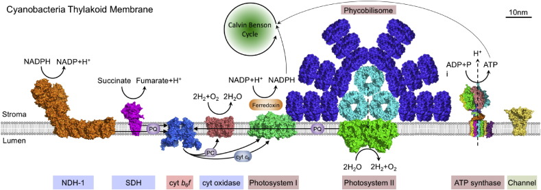
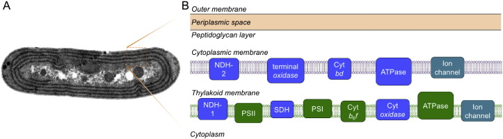
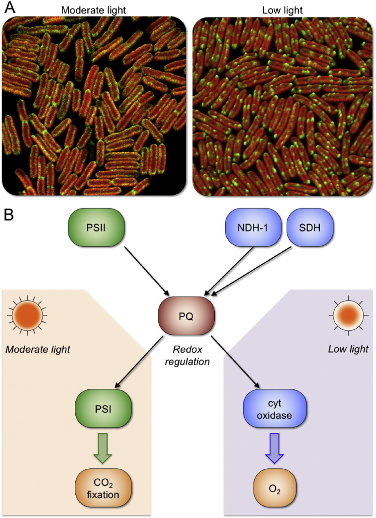
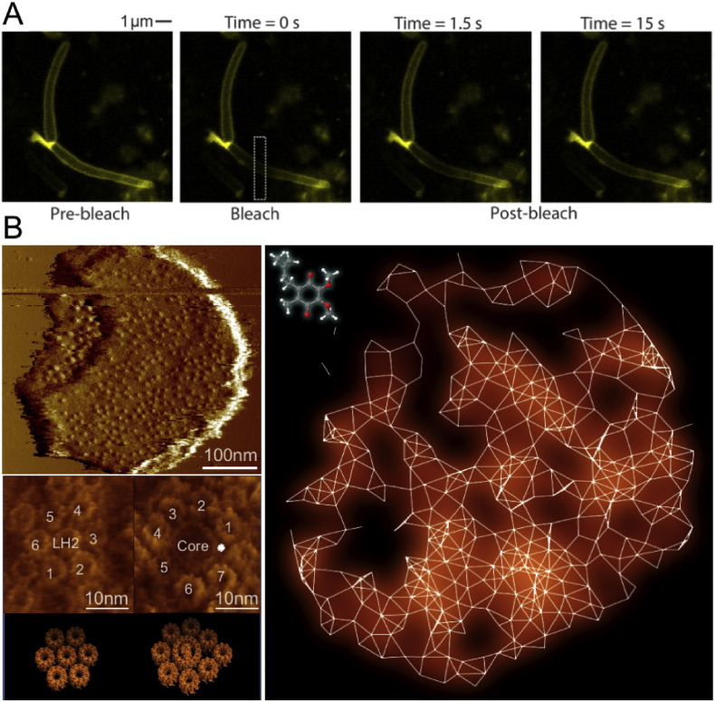
The cyanobacterial thylakoid membrane represents a system that can carry out both oxygenic photosynthesis and respiration simultaneously. The organization, interactions and mobility of components of these two electron transport pathways are indispensable to the biosynthesis of thylakoid membrane modules and the optimization of bioenergetic electron flow in response to environmental changes. These are of fundamental importance to the metabolic robustness and plasticity of cyanobacteria. This review summarizes our current knowledge about the distribution and dynamics of electron transport components in cyanobacterial thylakoid membranes. Global understanding of the principles that govern the dynamic regulation of electron transport pathways in nature will provide a framework for the design and synthetic engineering of new bioenergetic machinery to improve photosynthesis and biofuel production. This article is part of a Special Issue entitled: Organization and dynamics of bioenergetic systems in bacteria, edited by Conrad Mullineaux.

Keywords: Cyanobacteria; Electron transport; Membrane protein; Photosynthesis; Protein distribution; Protein dynamics; Respiration; Supramolecular complex; Thylakoid membrane.


Figures

Fig. 1
Fig. 1
Schematic model of cyanobacterial thylakoid membrane (based on knowledge of Synechocystis 6803 thylakoids), showing the interplay of photosynthetic and respiratory electron transport component in the same membrane. Photosynthetic electron transfer complexes include phycobilisome, PSII and PSI, cyt b6f and ATPase. The presence of phycobilisome–photosystem supercomplex in vivo has been identified , . Complexes specific for respiratory electron transport chain are NDH-1, SDH and cyt oxidase. Some components, such as the cyt b6f, PQ and PC are shared by both electron transport pathways. There are also potassium channel proteins in the thylakoid membrane. Arrows indicate the electron transduction reactions. Abbreviations: ADP — adenosine diphosphate, ATP — adenosine triphosphate, cyt b6f — cytochrome b6f, cyt c6 — cytochrome c6, cyt oxidase — cytochrome oxidase, NADP(H) — nicotinamide-adenine dinucleotide phosphate (reduced form), NDH-1 — type 1 NADPH dehydrogenase, PC — plastocyanin, PQ — plastoquinone, SDH — succinate dehydrogenase. The protein structures are achieved from PDB database: allophycocyanin, PDB ID: 1KN1; NDH-1, based on the Complex I structure from Thermus thermophilus, PDB ID: 4HEA; cyt b6f, PDB ID: 4H13; cyt oxidase, PDB ID: 1OCO; potassium channel protein, based on the Magnetospirillum magnetotacticum KirBac3.1 potassium channel crystal structure, PDB ID: 1XL4; phycocyanin, PDB ID: 3O18; PSI, PDB ID: 1JB0; PSII, PDB ID: 3WU2; and SDH, based on the E. coli SDH crystal structures, PDB ID: 1NEK.
Fig. 2
Fig. 2
Model of cyanobacterial membrane system and the distribution of electron transport complexes in the membranes. Left, thin-section electron microscopy image of a Synechococcus 7942 cell. The thylakoid membranes of Synechococcus are organized in a series of regular, concentric layers along the length of the cell. Right, distribution of the major components of cyanobacterial electron transport pathways in the cytoplasmic and thylakoid membranes. Respiratory electron transport components (blue) are located in both cytoplasmic and thylakoid membranes. The thylakoid membrane houses complexes from both photosynthetic (green) and respiratory electron transport chains. Abbreviations: ATPase — ATP synthase, cyt bd — cytochrome bd oxidase, NDH-1 and -2 — type 1 and II NADPH dehydrogenase, terminal oxidase — cytochrome terminal oxidase.
Fig. 3
Fig. 3
Regulation of electron transport pathways. A, Confocal microscopy images show different localization of functional NDH-1 complexes in Synechococcus 7942 grown in moderate light (60 μE m− 2 s− 1) and low light (6 μE m− 2 s− 1). The fluorescence of green fluorescence proteins (shown in green) revealed the distribution of NDH-1 complexes, whereas chlorophyll fluorescence (shown in red) indicates the location of thylakoid membranes. The reorganization of NDH-1 complexes in thylakoid membranes highly correlates with the direction of electron flow to respiratory pathway or photosynthetic pathway. For details, see . B, Model of the regulation of electron transport pathways in cyanobacterial thylakoid membranes. Arrows indicate the directions of electron flow. Complexes related to photosynthesis are shown in green and respiratory components are indicated in blue. Different light intensities could trigger the variation of the redox state of PQ pool, and thereby lead to distinct pathways of electron transport.
Fig. 4
Fig. 4
Mobility and diffusion pathways of electron carriers in the cellular membrane. A, Fluorescence recovery after photobleaching of living E. coli cells incubated with NBDHA-Q indicated the ubiquinone mobility in bacterial membrane. For details, see . B, Network of quinone pathways within the entire intracytoplasmic photosynthetic membranes from purple photosynthetic bacteria. Overview atomic force microscopy images and molecular-resolution images of typical photosynthetic protein assemblies of the native photosynthetic membranes from Rhodospirillum photometricum revealed specific protein organization and crowding in photosynthetic membranes, which are the fundamental basis of the long-range quinone pathways in cellular membranes. For details, see .

References

    1. Schopf J.W., Packer B.M. Early Archean (3.3-billion to 3.5-billion-year-old) microfossils from Warrawoona Group, Australia. Science. 1987;237:70–73. - PubMed
    1. Vermaas W.F. Nature Publishing Group; London: 2001. Photosynthesis and Respiration in Cyanobacteria, in: Encyclopedia of Life Sciences; pp. 245–251.
    1. Mullineaux C.W. Co-existence of photosynthetic and respiratory activities in cyanobacterial thylakoid membranes. Biochim. Biophys. Acta. 2014;1837:503–511. - PubMed
    1. DeRuyter Y.S., Fromme P. Molecular structure of the photosynthetic apparatus. In: Herrero A., Flores E., editors. The Cyanobacteria: Molecular Biology, Genomics, and Evolution. Caister Academic Press; Norfolk, UK: 2008. pp. 217–270.
    1. Adir N. Elucidation of the molecular structures of components of the phycobilisome: reconstructing a giant. Photosynth. Res. 2005;85:15–32. - PubMed

Publication types

MeSH terms

Substances

LinkOut - more resources