. 2016:29:59-68.
doi: 10.1007/8904_2015_495. Epub 2015 Dec 1.

Continual Low-Dose Infusion of Sulfamidase Is Superior to Intermittent High-Dose Delivery in Ameliorating Neuropathology in the MPS IIIA Mouse Brain

Affiliations

Continual Low-Dose Infusion of Sulfamidase Is Superior to Intermittent High-Dose Delivery in Ameliorating Neuropathology in the MPS IIIA Mouse Brain

Helen Beard et al. JIMD Rep. 2016.

Abstract

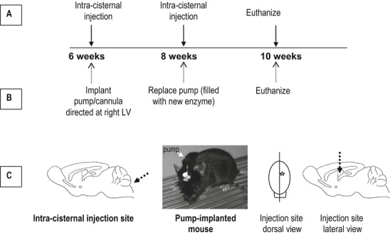
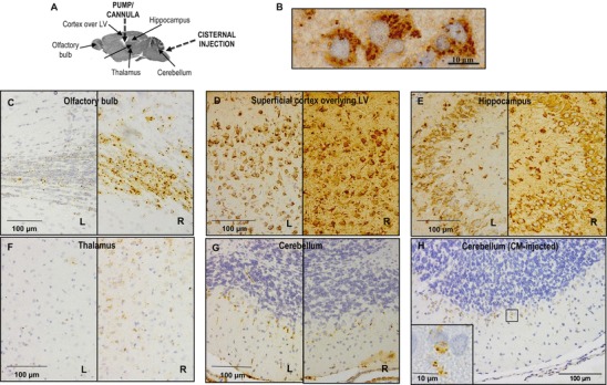
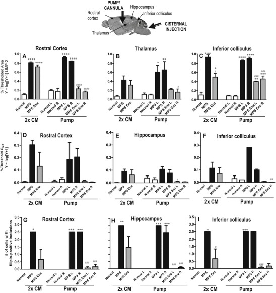
Mucopolysaccharidosis IIIA (MPS IIIA) is a neurodegenerative lysosomal storage disorder characterised by progressive loss of learned skills, sleep disturbance and behavioural problems. Reduced activity of lysosomal sulfamidase results in accumulation of heparan sulfate and secondary storage of glycolipids in the brain. Intra-cisternal sulfamidase infusions reduce disease-related neuropathology; however, repeated injections may subject patients to the risk of infection and tissue damage so alternative approaches are required. We undertook a proof-of-principle study comparing the ability of slow/continual or repeat/bolus infusion to ameliorate neuropathology in MPS IIIA mouse brain. Six-week-old MPS IIIA mice were implanted with subcutaneously located mini-osmotic pumps filled with recombinant human sulfamidase (rhSGSH) or vehicle, connected to lateral ventricle-directed cannulae. Pumps were replaced at 8 weeks of age. Additional MPS IIIA mice received intra-cisternal bolus infusions of the same amount of rhSGSH (or vehicle), at 6 and 8 weeks of age. Unaffected mice received vehicle via each strategy. All mice were euthanised at 10 weeks of age and the brain was harvested to assess the effect of treatment on neuropathology. Mice receiving pump-delivered rhSGSH exhibited highly significant reductions in lysosomal storage markers (lysosomal integral membrane protein-2, GM3 ganglioside and filipin-positive lipids) and neuroinflammation (isolectin B4-positive microglia, glial fibrillary acidic protein-positive astroglia). MPS IIIA mice receiving rhSGSH via bolus infusion displayed reductions in these markers, but the effectiveness of the strategy was inferior to that seen with slow/pump-based delivery. Continual low-dose infusion may therefore be a more effective strategy for enzyme delivery in MPS IIIA.


Figures

Fig. 1
Fig. 1
Experimental plan. Mice received either (a) bolus injection of 100 μg recombinant human sulfamidase (rhSGSH) via the cisterna magna (CM) at each of 6 and 8 weeks of age, with euthanasia at 10 weeks of age, or (b) implantation of a mini-osmotic pump and cannula, directed at the right lateral ventricle (LV) at 6 weeks of age. The pump was removed and replaced at 8 weeks of age and mice were euthanised at 10 weeks of age. During the experimental period, the pump delivered rhSGSH (1.2 μg/μL) at 0.25 μL/h, delivering 100 μg over each 2-week period. (c) Illustrates the injection region in each treatment strategy and shows a photo of a pump-/cannula-implanted mouse
Fig. 2
Fig. 2
Immunohistochemical localisation of rhSGSH in regions shown in (a) within the pump-/cannula-implanted (bg) and the cisternally injected (h) MPS IIIA mouse brain at 10 weeks of age. Staining in both left (L) and right (R) hemispheres of brain is shown for the cannulae-implanted mice (cg). The photo in (b) shows a high power view of the immunopositive puncta seen in the cerebral cortex of a pump-/cannula-implanted MPS IIIA mouse. The staining is presumptively in endo-/lysosomes
Fig. 3
Fig. 3
Quantification of immunohistochemical staining of lysosomal integral membrane protein-2 (LIMP-2; ac) and GM3 (df) and semi-quantification of filipin-stained inclusions (gi) in 10-week-old MPS IIIA and unaffected mouse brain following cisternal bolus (CM) or pump-delivered enzyme treatment. The location of the brain regions analysed with respect to the injection sites is shown in the sagittal brain section (*p < 0.05, **p < 0.01, ***p < 0.001, ****p < 0.0001 cf. Normal; #p < 0.05, ##p < 0.01, ###p < 0.001, ###p < 0.0001 cf. MPS vehicle)
Fig. 4
Fig. 4
Quantification of immunohistochemical staining of isolectin B4 (activated microglia; ac) and glial fibrillary acidic protein (GFAP; activated astrocytes; df) in 10-week-old MPS IIIA and unaffected mouse brain. The location of the brain regions analysed, with respect to the injection sites, is shown in the sagittal section of brain (*p < 0.05, **p < 0.01, ***p < 0.001, ****p < 0.0001 cf. Normal; #p < 0.05, ##p < 0.01, ###p < 0.001, ###p < 0.0001 cf. MPS vehicle)

References

    1. Chang M, Cooper JD, Sleat DE, et al. Intraventricular enzyme replacement improves disease phenotypes in a mouse model of late infantile neuronal ceroid lipofuscinosis. Mol Ther. 2008;16:649–656. doi: 10.1038/mt.2008.9. - DOI - PubMed
    1. Crawley AC, Gliddon BL, Auclair D, et al. Characterization of a C57BL/6 congenic mouse strain of mucopolysaccharidosis type IIIA. Brain Res. 2006;1104:1–17. doi: 10.1016/j.brainres.2006.05.079. - DOI - PubMed
    1. Crawley AC, Marshall N, Beard H, et al. Enzyme replacement reduces neuropathology in MPS IIIA dogs. Neurobiol Dis. 2011;43:422–434. doi: 10.1016/j.nbd.2011.04.014. - DOI - PubMed
    1. Dodge JC, Clarke J, Treleaven CM, et al. Intracerebroventricular infusion of acid sphingomyelinase corrects CNS manifestations in a mouse model of Niemann–Pick A disease. Exp Neurol. 2009;215:349–357. doi: 10.1016/j.expneurol.2008.10.021. - DOI - PubMed
    1. Gliddon BL, Hopwood JJ. Enzyme-replacement therapy from birth delays the development of behavior and learning problems in mucopolysaccharidosis type IIIA mice. Pediatr Res. 2004;56:65–72. doi: 10.1203/01.PDR.0000129661.40499.12. - DOI - PubMed

LinkOut - more resources