. 2015 Dec 1:6:236.
doi: 10.1186/s13287-015-0233-8.

A robust potency assay highlights significant donor variation of human mesenchymal stem/progenitor cell immune modulatory capacity and extended radio-resistance

Affiliations

A robust potency assay highlights significant donor variation of human mesenchymal stem/progenitor cell immune modulatory capacity and extended radio-resistance

Nina Ketterl et al. Stem Cell Res Ther. .

Abstract

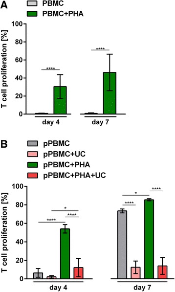
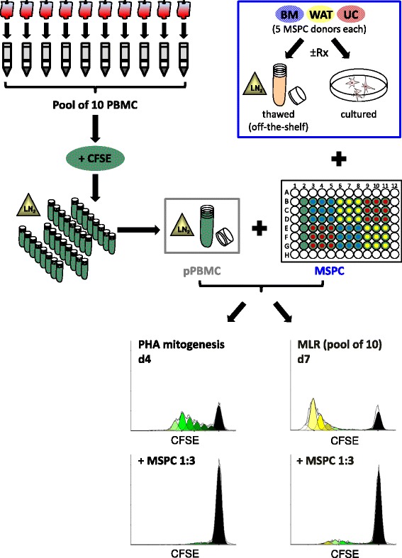
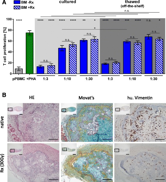
The inherent immunomodulatory capacity of mesenchymal stem/progenitor cells (MSPCs) encouraged initiation of multiple clinical trials. Release criteria for therapeutic MSPCs cover identity, purity and safety but appropriate potency assessment is often missing. Reports on functional heterogeneity of MSPCs created additional uncertainty regarding donor and organ/source selection. We established a robust immunomodulation potency assay based on pooling responder leukocytes to minimize individual immune response variability. Comparing various MSPCs revealed significant potency inconsistency and generally diminished allo-immunosuppression compared to dose-dependent inhibition of mitogenesis. Gamma-irradiation to block unintended MSPC proliferation did not prohibit chondrogenesis and osteogenesis in vivo, indicating the need for alternative safety strategies.


Figures

Fig. 1
Fig. 1
Individual or pooled donor polyclonal T-cell proliferation. a Mean ± SD proliferation of five random single donor buffy coat-derived CFSE-labeled peripheral blood mononuclear cells (PBMCs) in the absence (grey bars) or presence of phytohemagglutinin (+PHA, green bars) after 4 or 7 days indicating significant donor variation. b Simultaneous inhibition of mitogenic and alloimmunogenic T-cell proliferation by umbilical cord (UC)-derived MSPCs was tested. CFSE pre-labeled cryopreserved pooled PBMCs (pPBMCs) were seeded in the absence of MSPCs without (grey bars) or with PHA (green bars) or in the presence of UC-MSPC without (light red bars; inhibition of allo-MLR) or with PHA (red bars; inhibition of additional mitogenesis) at an MSPC:PBMC ratio of 1:3. The time course of proliferation between days 4 to 7 is shown in Additional file 2 (Figure S1C). Mean ± SD results from representative experiments (n = 5 in a and b). Significant differences are indicated (*p < 0.05; **p < 0.01; ***p < 0.001; ****p < 0.0001)
Fig. 2
Fig. 2
Illustrated potency assay strategy. Ten randomly obtained buffy coats from healthy donors can be processed in parallel to isolate approximately 1 × 109 peripheral blood mononuclear cells (PBMCs) per donor, pooled, labeled with carboxyfluorescein (CFSE) and cryopreserved in appropriate aliquots (e.g., 200 aliquots of 1 × 107 pre-labeled pooled PBMCs (pPBMC)) for subsequent off-the-shelf use as responder cells in the potency assay. Individual mesenchymal stem/progenitor cells (MSPC) from donor or organ origin of choice (e.g., bone marrow (BM), white adipose tissue (WAT), umbilical cord (UC); color code corresponding to Fig. 1) can be tested off-the-shelf or after rescue culture (with or without gamma irradiation (±Rx)) to test their potency to inhibit mitogen (e.g., PHA)-driven pPBMC proliferation until day 4 (d4), or to inhibit the allogeneic mixed lymphocyte reaction (MLR) of the same pPBMC batch until day 7 (d7). Representative CD3+ T-cell proliferation kinetics (Modfit analysis) in the absence (top histograms), or presence of regulatory MSPCs (bottom histograms) indicating maximum number of proliferated populations (top histograms) and the effect of MSPC-mediated inhibition of T-cell proliferation (bottom histograms) are shown
Fig. 3
Fig. 3
Pooled pre-labeled PBMC and pooled reference MSPC make a robust assay format to readout inhibition of T-cell proliferation in an off-the-shelf potency assay. a, b Pooled carboxyfluorescein pre-labeled random donor peripheral blood mononuclear cell (pPBMC) aliquots seeded in triplicate show a highly significant mitogen-induced proliferation (phytohemagglutinin (PHA); green bars) compared to minimum proliferation of the unstimulated pPBMC seeded off-the-shelf in the absence of PHA (dark grey bar) at day 4 (d4). Triplicates of pMSPC composed of cells from five each random donors (D1D15) of bone marrow (BM; blue bars), white adipose tissue (WAT; yellow bars) and umbilical cord (UC; red bars) were used as an organotypic MSPC reference (grey areas) to determine organ-specific highly significant inhibition of mitogen-induced T-cell proliferation compared to individual a cultured or b freshly thawed individual MSPC from five donors per organ. c, d At day 7 (d7), the potent allo-response of pPBMC (grey bars; for time course titration see Fig. 1b, c) in the absence of external stimulation was significantly inhibited by some but not all individual MSPCs compared to the organotypic reference pMSPC (grey areas; same donors and identical color code as in a, b). Differences between c cultured and d freshly thawed individual or pooled MSPCs were more prominent compared to the inhibition of mitogenesis (in a, b). Significant inhibition of day 4 mitogen-induced (a, b) and day 7 mixed leukocyte reaction (MLR)-induced (c, d) pooled T-cell proliferation is indicated at the right margin of the graphs; significance of individual donor comparison is indicated by vertical lines. Mean ± SD results at an MSPC:PBMC ratio of 1:3 are shown. *p < 0.05; **p < 0.01 ***p < 0.001; ****p < 0.0001
Fig. 4
Fig. 4
Irradiated MSPC maintain their immunomodulatory potency in vitro and their differentiation capacity in vivo. a Direct comparison of the inhibition of phytohemagglutinin (PHA)-induced T-cell proliferation (green bar) by non-irradiated bone marrow (BM)-MSPCs (-Rx; blue bars) versus 30 Gy irradiated BM-MSPCs (+Rx; hatched blue bars) immediately after thawing (off-the-shelf use; dark grey area) or after a 3-day rescue culture (light grey area) showed no significant difference at the three ratios as indicated (*p < 0.05; **p < 0.01; ***p < 0.001; ****p < 0.0001). Grey bar shows mean ± SD of unstimulated pooled T-cell proliferation. One representative experiment out of two is shown. b Representative histologic analysis of ectopic ossicles derived from native (non-irradiated, upper pictures) and irradiated (Rx; 30 Gy, lower pictures) BM-MSPC (n = 6 per group) 6 weeks after subcutaneous transplantation into immunocompromized NSG recipient mice. Bone formation via a vascularized cartilage intermediate was evident in hematoylin and eosin (HE; left panels) as well as Movat’s pentachrome (Movat; middle panels) staining. Vimentin staining (right panels) indicating persistence of reticular stromal cells (MSPCs) within the ectopic ossicles which showed infiltration by (human (hu.) Vimentin-negative) murine hematopoiesis as described previously only for native (non-irradiated) BM-MSPCs [27]. Scale bars are 100 μm in main histophotographs and 1 mm in inserts (showing overview of a section through the entire ossicle; dotted rectangles indicate the regions from where the magnified main pictures were derived). n.s. Not significant, pPBMC Pooled peripheral mononuclear cell

References

    1. Caplan AI, Correa D. The MSC: an injury drugstore. Cell Stem Cell. 2011;9(1):11–5. doi: 10.1016/j.stem.2011.06.008. - DOI - PMC - PubMed
    1. Bianco P, Cao X, Frenette PS, Mao JJ, Robey PG, Simmons PJ, et al. The meaning, the sense and the significance: translating the science of mesenchymal stem cells into medicine. Nat Med. 2013;19(1):35–42. doi: 10.1038/nm.3028. - DOI - PMC - PubMed
    1. Mimeault M, Hauke R, Batra SK. Stem cells: a revolution in therapeutics-recent advances in stem cell biology and their therapeutic applications in regenerative medicine and cancer therapies. Clin Pharmacol Ther. 2007;82(3):252–64. doi: 10.1038/sj.clpt.6100301. - DOI - PubMed
    1. Strunk D. Endothelial progenitor cells: quod erat demonstrandum? Curr Pharm Des. 2011;17(30):3245–51. doi: 10.2174/138161211797904127. - DOI - PubMed
    1. Caimi PF, Reese J, Lee Z, Lazarus HM. Emerging therapeutic approaches for multipotent mesenchymal stromal cells. Curr Opin Hematol. 2010;17(6):505–13. doi: 10.1097/MOH.0b013e32833e5b18. - DOI - PMC - PubMed

Publication types