. 2016 Jan:90:252-260.
doi: 10.1016/j.freeradbiomed.2015.11.026. Epub 2015 Nov 24.

Nitro-oleic acid modulates classical and regulatory activation of macrophages and their involvement in pro-fibrotic responses

Affiliations

Nitro-oleic acid modulates classical and regulatory activation of macrophages and their involvement in pro-fibrotic responses

Gabriela Ambrozova et al. Free Radic Biol Med. 2016 Jan.

Abstract

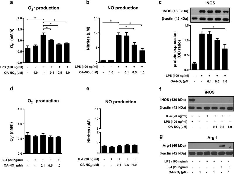
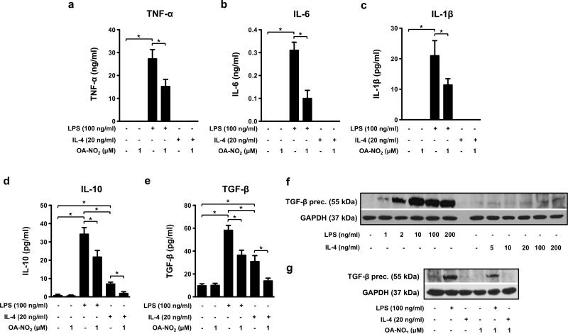
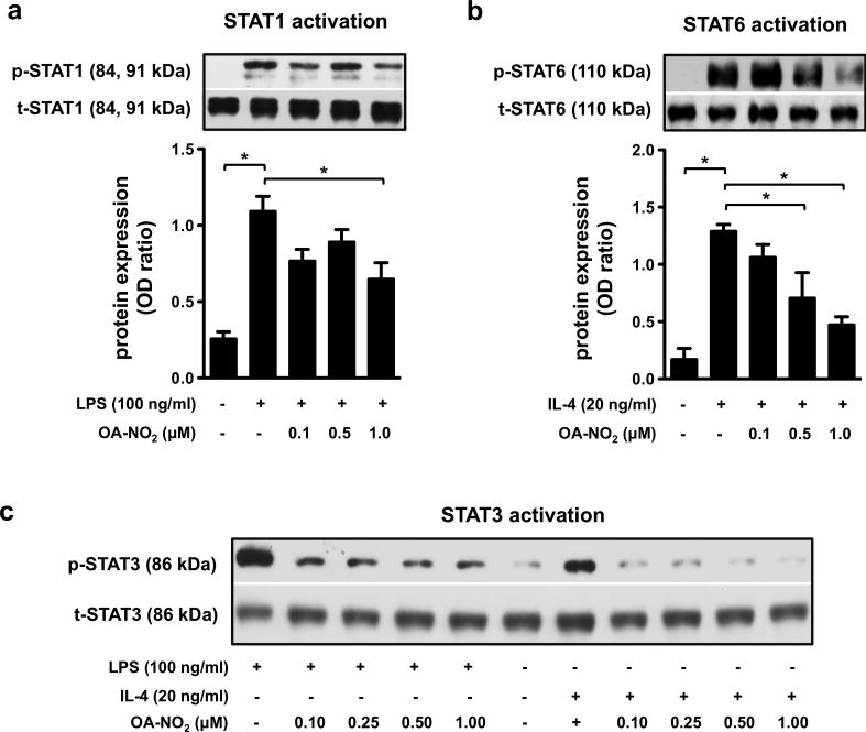
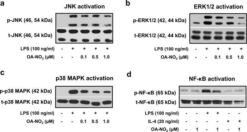
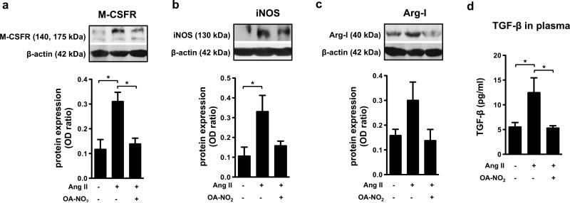
Inflammation is an immune response triggered by microbial invasion and/or tissue injury. While acute inflammation is directed toward invading pathogens and injured cells, thus enabling tissue regeneration, chronic inflammation can lead to severe pathologies and tissue dysfunction. These processes are linked with macrophage polarization into specific inflammatory "M1-like" or regulatory "M2-like" subsets. Nitro-fatty acids (NO2-FAs), produced endogenously as byproducts of metabolism and oxidative inflammatory conditions, may be useful for treating diseases associated with dysregulated immune homeostasis. The goal of this study was to characterize the role of nitro-oleic acid (OA-NO2) in regulating the functional specialization of macrophages induced by bacterial lipopolysaccharide or interleukin-4, and to reveal specific signaling mechanisms which can account for OA-NO2-dependent modulation of inflammation and fibrotic responses. Our results show that OA-NO2 inhibits lipopolysaccharide-stimulated production of both pro-inflammatory and immunoregulatory cytokines (including transforming growth factor-β) and inhibits nitric oxide and superoxide anion production. OA-NO2 also decreases interleukin-4-induced macrophage responses by inhibiting arginase-I expression and transforming growth factor-β production. These effects are mediated via downregulation of signal transducers and activators of transcription, mitogen-activated protein kinase and nuclear factor-кB signaling responses. Finally, OA-NO2 inhibits fibrotic processes in an in vivo model of angiotensin II-induced myocardial fibrosis by attenuating expression of α-smooth muscle actin, systemic transforming growth factor-β levels and infiltration of both "M1-" and "M2-like" macrophage subsets into afflicted tissue. Overall, the electrophilic fatty acid derivative OA-NO2 modulates a broad range of "M1-" and "M2-like" macrophage functions and represents a potential therapeutic approach to target diseases associated with dysregulated macrophage subsets.

Keywords: Fibrosis; Inflammation; Macrophage functional specialization; Macrophages; Nitro-fatty acids; Nitro-oleic acid.


Figures

Figure 1
Figure 1. OA-NO2 downregulates O2.- and NO production, iNOS and arginase-I expression in “M1-” and “M2-like” RAW 264.7 macrophages
Cells were treated with different concentrations of OA-NO2 (0.1, 0.5, 1.0 μM) and stimulated with LPS (100 ng/ml) or IL-4 (20 ng/ml). O2.- production was monitored for 4 h (n=6) (a, d). Production of NO (n=6) (b, e), expression of iNOS (n=3) (c, f) and arginase-I (g) was detected in macrophages incubated for 24 h. The pictures represent one of three individual experiments. A *p value of less than 0.05 was considered significant when evaluating differences between the individual bars and positive control (LPS- and IL-4-treated cells) or between two individual bars, respectively.
Figure 2
Figure 2. OA-NO2 affects the cytokine profile in “M1-” and “M2-like” RAW 264.7 macrophages
Cells were treated with OA-NO2 (1.0 μM) and stimulated with LPS (100 ng/ml) or IL-4 (20 ng/ml) for 24 h. TNF-α (a), IL-6 (b), IL-1β (c), IL-10 (d) and TGF-β (e) production was measured in cell supernatants (n=6-8). The expression of TGF-β precursor was monitored in RAW 264.7 macrophages treated with different concentrations of LPS (1-200 ng/ml), IL-4 (5-200 ng/ml), and/or OA-NO2 (1uM) (f). The pictures represent one of three individual experiments. A *p value of less than 0.05 was considered significant when evaluating differences between the individual bars and positive control (LPS- and IL-4-treated cells) or between two individual bars, respectively.
Figure 3
Figure 3. OA-NO2 downregulates the LPS- and IL-4-induced activation of STAT1, STAT3, and STAT6 in “M1-” and “M2-like” RAW 264.7 macrophages
The expression and phosphorylation of STATs was detected in RAW 264.7 cells treated with different concentrations of OA-NO2 (0.1, 0.25, 0.5, 1.0 μM) and stimulated with LPS (100 ng/ml) (a, c) or IL-4 (20 ng/ml) (b, c) for 1 h. Expression of p-STAT1/t-STAT1 (a), p-STAT6/t-STAT6 (b), and p-STAT3/t-STAT3 (c) was detected in both “M1-” and “M2-like” macrophages (n=3). The pictures represent one of three individual experiments. A *p value of less than 0.05 was considered significant when evaluating differences between the individual bars and positive control (LPS- and IL-4-treated cells) or between two individual bars, respectively.
Figure 4
Figure 4. OA-NO2 regulates the activation of MAPKs and NF-κB in “M1-like” RAW 264.7 macrophages
Cells were treated with OA-NO2 (0.1, 0.5, 1.0 μM) and exposed to LPS (100 ng/ml) or IL-4 (20 ng/ml). Phosphorylation of MAPKs (a-c) was detected 1 h after cell stimulation with LPS or IL-4. Expression and activation of NF-κB was monitored in both “M1-” and “M2-like” macrophage subpopulations treated for 30 min (d). The pictures represent one of three individual experiments.
Figure 5
Figure 5. OA-NO2 decreases the Ang II-induced fibrotic processes in heart tissue
The expression of M-CSFR (a), iNOS (b), and arginase-I (c) was detected in heart tissue of C57BL/6J mice treated for 2 weeks with Ang II (1.5 ng/g/min) and OA-NO2 (6 mg/kg) via subcutaneously implanted osmotic minipumps. The concentration of TGF-β was determined in plasma samples (n=6-9) (d). The pictures represent one of several individual experiments (n=6-9). A *p value of less than 0.05 was considered significant when evaluating differences between the individual bars and positive control (Ang II-treated mice).

References

    1. Nahrendorf M, Swirski FK. Monocyte and macrophage heterogeneity in the heart. Circulation research. 2013;112:1624–1633. - PMC - PubMed
    1. Biswas SK, Chittezhath M, Shalova IN, Lim JY. Macrophage polarization and plasticity in health and disease. Immunologic research. 2012;53:11–24. - PubMed
    1. Lawrence T, Natoli G. Transcriptional regulation of macrophage polarization: enabling diversity with identity. Nature reviews. Immunology. 2011;11:750–761. - PubMed
    1. Eguchi J, Kong X, Tenta M, Wang X, Kang S, Rosen ED. Interferon regulatory factor 4 regulates obesity-induced inflammation through regulation of adipose tissue macrophage polarization. Diabetes. 2013;62:3394–3403. - PMC - PubMed
    1. Ohmori Y, Hamilton TA. Requirement for STAT1 in LPS-induced gene expression in macrophages. Journal of leukocyte biology. 2001;69:598–604. - PubMed

Publication types