Case Reports
. 2015 Dec 1:7:113.
doi: 10.1186/s13073-015-0235-2.

Clinical metagenomic identification of Balamuthia mandrillaris encephalitis and assembly of the draft genome: the continuing case for reference genome sequencing

Affiliations
Case Reports

Clinical metagenomic identification of Balamuthia mandrillaris encephalitis and assembly of the draft genome: the continuing case for reference genome sequencing

Alexander L Greninger et al. Genome Med. .

Erratum in

Abstract

Background: Primary amoebic meningoencephalitis (PAM) is a rare, often lethal, cause of encephalitis, for which early diagnosis and prompt initiation of combination antimicrobials may improve clinical outcomes.

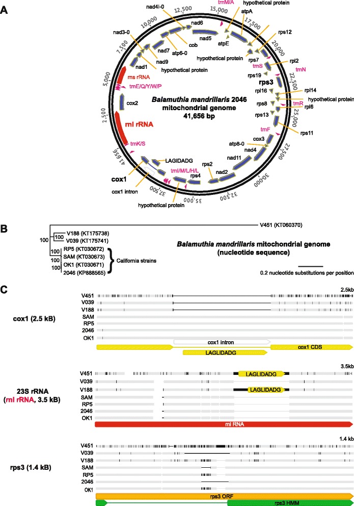
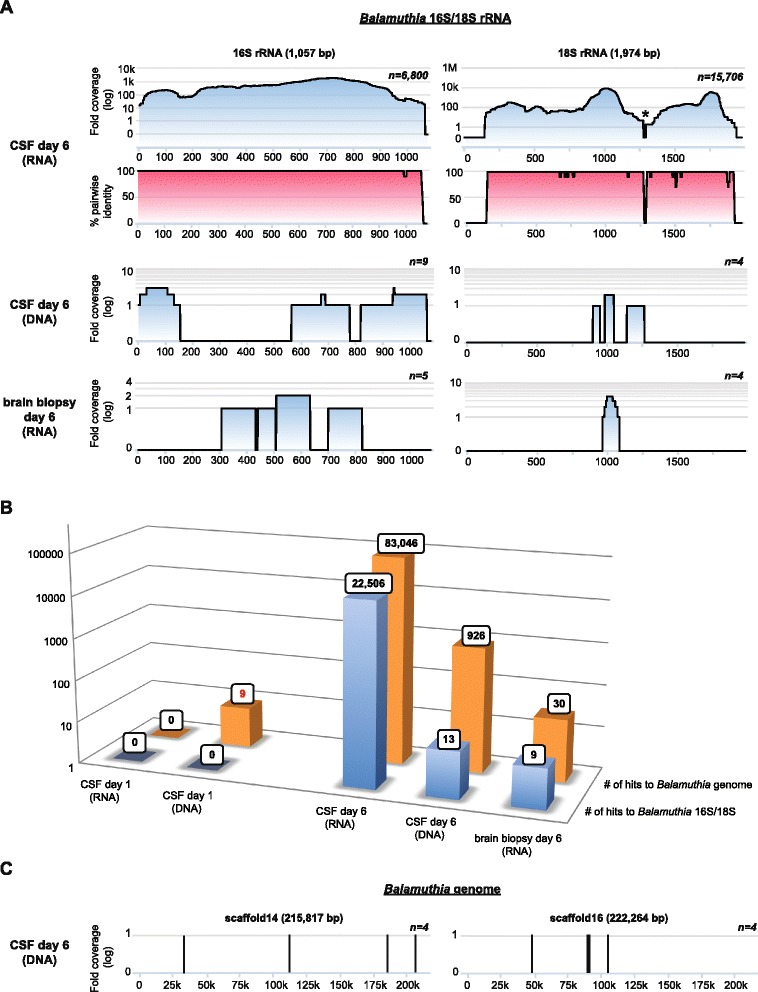
Methods: In this study, we sequenced a full draft assembly of the Balamuthia mandrillaris genome (44.2 Mb in size) from a rare survivor of PAM, and recovered the mitochondrial genome from six additional Balamuthia strains. We also used unbiased metagenomic next-generation sequencing (NGS) and SURPI bioinformatics analysis to diagnose an ultimately fatal case of Balamuthia mandrillaris encephalitis in a 15-year-old girl.

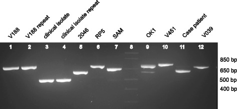
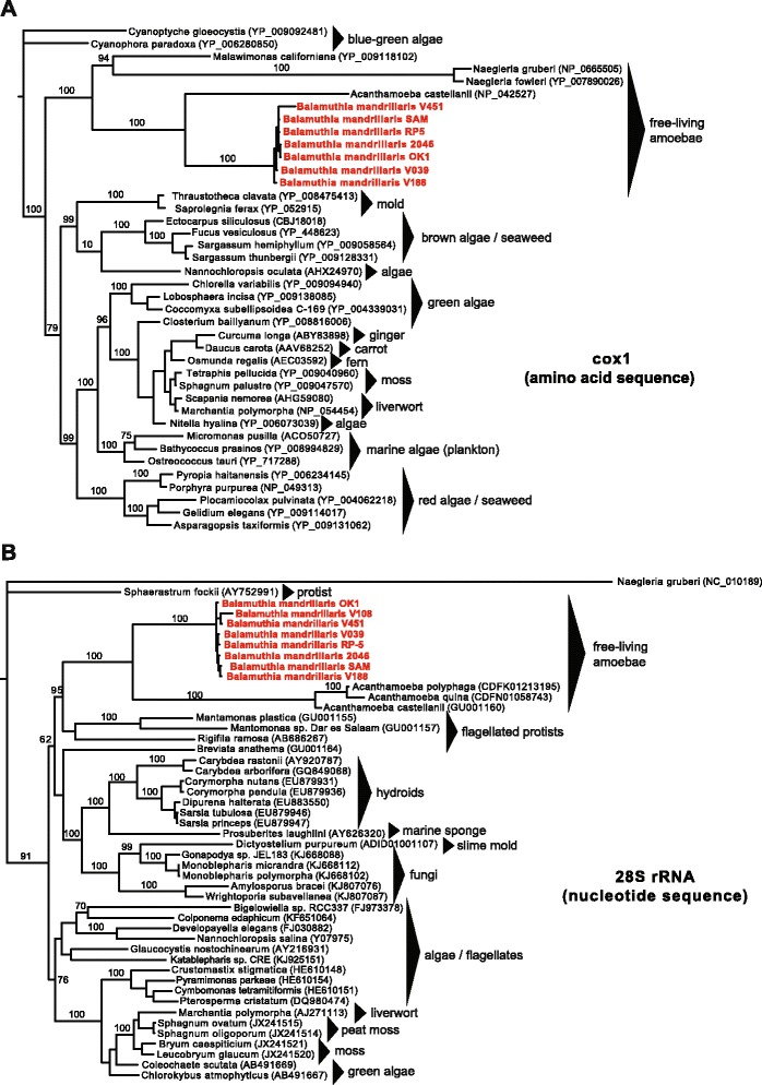
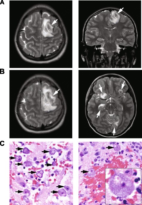
Results and discussion: Comparative analysis of the mitochondrial genome and high-copy number genes from six additional Balamuthia mandrillaris strains demonstrated remarkable sequence variation, and the closest Balamuthia homologs corresponded to other amoebae, hydroids, algae, slime molds, and peat moss. Real-time NGS testing of hospital day 6 CSF and brain biopsy samples detected Balamuthia on the basis of high-quality hits to 16S and 18S ribosomal RNA sequences present in the National Center for Biotechnology Information (NCBI) nt reference database. The presumptive diagnosis of PAM by visualization of amoebae on brain biopsy histopathology and NGS analysis was subsequently confirmed at the US Centers for Disease Control and Prevention (CDC) using a Balamuthia-specific PCR assay. Retrospective analysis of a day 1 CSF sample revealed that more timely identification of Balamuthia by metagenomic NGS, potentially resulting in a better clinical outcome, would have required availability of the complete genome sequence.

Conclusions: These results underscore the diverse evolutionary origins of Balamuthia mandrillaris, provide new targets for diagnostic assay development, and will facilitate further investigations of the biology and pathogenesis of this eukaryotic pathogen. The failure to identify PAM from a day 1 sample without a fully sequenced Balamuthia genome in the database highlights the critical importance of whole-genome reference sequences for microbial detection by metagenomic NGS.


Figures

Fig. 1
Fig. 1
Sequencing and comparative phylogenetic analysis of the mitochondrial genome of Balamuthia mandrillaris. a Balamuthia mandrillaris 2046 mitochondrial genome. Annotation of the 41,656 bp genome was performed using RNAmmer, tRNAscan-SE, and Glimmer gene predictor, with all ORFs manually verified using BLASTx alignment. b Phylogenetic analysis of seven newly sequenced mitochondrial genomes from different strains of Balamuthia mandrillaris. An outgroup (for example, Acanthamoeba castellani) is not shown given the lack of gene synteny. Branch lengths are drawn proportionally to the number of nucleotide substitutions per position, and support values are shown for each node. c Differences in individual gene features (cox1, 23S rRNA, and rps3), among the 7 mitochondrial genomes, as detailed in the text. The mitochondrial cox1, 23S rRNA / rnl RNA, and rps3 genes are highlighted in boldface
Fig. 2
Fig. 2
PCR amplification of the Balamuthia rps3 mitochondrial gene. The variable length of the rps3 intron among eight different Balamuthia strains (seven newly sequenced mitochondrial genomes and the case patient) suggests that this gene may be an attractive target for development of a molecular genotyping assay. Lane 8 corresponds to the DNA ladder (faint appearance), while lanes 3 and 4 correspond to an additional clinical Balamuthia isolate whose mitochondrial genome was not sequenced
Fig. 3
Fig. 3
Phylogenetic trees of the mitochondrial cox1 protein and 28S rRNA gene reveal the close phylogenetic relationship between Balamuthia and Acanthamoeba. a Phylogeny of seven Balamuthia cox1 amino acid sequences along with the top complete sequence hits in NCBI nr ranked by BLASTp E-score. b Phylogeny of seven Balamuthia 23S rRNA nucleotide sequences along with the top complete sequence hits in NCBI nt ranked by BLASTn E-score. Sequences were aligned using MUSCLE and a phylogenetic tree constructed using MrBayes. Branch lengths are drawn proportionally to the number of nucleotide substitutions per position, and support values are shown for each node
Fig. 4
Fig. 4
MRI and histopathology from a 15-year-old patient with a fulminant acute encephalitis. a A hospital day (HD) 1 coronal T2-weighted MR image, demonstrating a hemorrhagic lesion with surrounding edema within the superior left frontal lobe (left panel, white arrow) and left occipital lobe (right panel, white arrow). b A HD 5 contrast-enhanced T1-weighted MR image, revealing enlargement of the pre-existing left frontal lobe lesion (left panel, white arrow), as well as interval development of numerous additional rim-enhancing lesions in multiple regions (right panel, white arrows). c 20× (left and right panels) and 100× fields of view (right panel, inset) of a brain biopsy specimen from the patient demonstrating numerous viable, large amoebae (black arrows), with abundant basophilic vacuolated cytoplasm, round central nuclei, and prominent nucleoli, consistent with Balamuthia mandrillaris. There were areas of extensive hemorrhagic necrosis accompanied by a polymorphic inflammatory cell infiltrate including neutrophils and eosinophils (right panel)
Fig. 5
Fig. 5
Identification of Balamuthia mandrillaris infection by metagenomic next-generation sequencing (NGS). a Coverage maps (blue gradient) and pairwise identity plots (magenta gradient) of two of the three available sequences from Balamuthia (16S/18S rRNA genes) in the NCBI nt reference database as of August 2015. Shown are coverage maps corresponding to day 6 DNA and RNA libraries from CSF and a day 6 mRNA library from brain biopsy. No hits to 16S and 18S Balamuthia sequences were seen from day 1 samples. The asterisk denotes an area with artificially low coverage after taxonomic classification of the NGS reads due to high conservation among eukaryotic sequences (for example, human, Balamuthia, and so on) within that region. b A bar graph of the number of species-specific NGS reads aligning to Balamuthia 16S/18S rRNA (blue) or the Balamuthia genome (orange) in day 1 or day 6 samples. Note that with the availability of the newly assembled 44 Mb Balamuthia genome, diagnosis of Balamuthia mandrillaris encephalitis at day 1 would have possible by detection of nine species-specific reads (red boldface). c Coverage maps of two large scaffolds, approximately 216 kB and 222 kB in size, from the Balamuthia draft genome, showing eight out of 926 hits to Balamuthia in the day 6 CSF DNA library that are identified by SURPI after the draft genome sequence is added to the reference database (versus only 13 hits previously)

References

    1. Visvesvara GS. Epidemiology of infections with free-living amebas and laboratory diagnosis of microsporidiosis. Mt Sinai J Med. 1993;60:283–8. - PubMed
    1. Matin A, Siddiqui R, Jayasekera S, Khan NA. Increasing importance of Balamuthia mandrillaris. Clin Microbiol Rev. 2008;21:435–48. doi: 10.1128/CMR.00056-07. - DOI - PMC - PubMed
    1. Schuster FL, Guglielmo BJ, Visvesvara GS. In-vitro activity of miltefosine and voriconazole on clinical isolates of free-living amebas: Balamuthia mandrillaris, Acanthamoeba spp., and Naegleria fowleri. J Eukaryot Microbiol. 2006;53:121–6. doi: 10.1111/j.1550-7408.2005.00082.x. - DOI - PubMed
    1. Visvesvara GS, Martinez AJ, Schuster FL, Leitch GJ, Wallace SV, Sawyer TK, et al. Leptomyxid ameba, a new agent of amebic meningoencephalitis in humans and animals. J Clin Microbiol. 1990;28:2750–6. - PMC - PubMed
    1. Schuster FL, Dunnebacke TH, Booton GC, Yagi S, Kohlmeier CK, Glaser C, et al. Environmental isolation of Balamuthia mandrillaris associated with a case of amebic encephalitis. J Clin Microbiol. 2003;41:3175–80. doi: 10.1128/JCM.41.7.3175-3180.2003. - DOI - PMC - PubMed

Publication types