. 2016 Jan 12;113(2):E172-81.
doi: 10.1073/pnas.1521674112. Epub 2015 Nov 30.

Functional screen identifies kinases driving prostate cancer visceral and bone metastasis

Affiliations

Functional screen identifies kinases driving prostate cancer visceral and bone metastasis

Claire M Faltermeier et al. Proc Natl Acad Sci U S A. .

Abstract

Mutationally activated kinases play an important role in the progression and metastasis of many cancers. Despite numerous oncogenic alterations implicated in metastatic prostate cancer, mutations of kinases are rare. Several lines of evidence suggest that nonmutated kinases and their pathways are involved in prostate cancer progression, but few kinases have been mechanistically linked to metastasis. Using a mass spectrometry-based phosphoproteomics dataset in concert with gene expression analysis, we selected over 100 kinases potentially implicated in human metastatic prostate cancer for functional evaluation. A primary in vivo screen based on overexpression of candidate kinases in murine prostate cells identified 20 wild-type kinases that promote metastasis. We queried these 20 kinases in a secondary in vivo screen using human prostate cells. Strikingly, all three RAF family members, MERTK, and NTRK2 drove the formation of bone and visceral metastasis confirmed by positron-emission tomography combined with computed tomography imaging and histology. Immunohistochemistry of tissue microarrays indicated that these kinases are highly expressed in human metastatic castration-resistant prostate cancer tissues. Our functional studies reveal the strong capability of select wild-type protein kinases to drive critical steps of the metastatic cascade, and implicate these kinases in possible therapeutic intervention.

Keywords: bone metastasis; kinases; metastasis; prostate cancer.


Conflict of interest statement

The authors declare no conflict of interest.

Figures

Fig. 1.
Fig. 1.
Schematic summary of the screen for metastasis-promoting kinases. One hundred twenty-five candidate kinases were identified from a combination of genomic/transcriptomic, phosphoproteomic, and literature data. The primary screen entailed expressing all 125 kinases individually in a murine cell line followed by tail vein injection of cells into recipient mice. Twenty kinases strongly promoted lung colonization in vivo. The 20 kinases identified in the primary screen were subjected to a secondary in vivo screen using human prostate cells. Five kinases promoted bone and visceral metastasis in the human cell context.
Fig. S1.
Fig. S1.
Lentivirus-mediated overexpression of V5-tagged kinases. (A) Full-length kinases were cloned into the FU-R1-R2-V5-SV40-Blasti-CGW lentiviral vector shown. R1 and R2 represent recombination sites required for recombination-based Gateway cloning. LTR, Long-terminal repeat. (B) Western blot showing expression of selected kinases in 293t cells detected by a V5 antibody. The molecular mass of each kinase is indicated in parentheses.
Fig. S2.
Fig. S2.
SrcY529F promotes lung colonization when overexpressed in murine prostate cells. (A) Experimental design to demonstrate that expression of mutationally activated kinase SrcY529F in Cap8 cells promotes lung colonization. (B) Bright-field images of lungs removed from mice 3 wk after being injected with Cap8-SrcY529F cells. (Scale bars, 5 mm.)
Fig. 2.
Fig. 2.
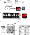
In vivo screen of 125 candidate kinases identifies 20 kinases with metastasis-promoting ability when expressed in murine prostate cells. (A) Schematic diagram of the screen testing the metastatic ability of 125 kinases. Kinases were expressed individually in Cap8 cells, pooled into groups of five kinases (each with a different molecular weight), and injected into the tail vein of CB17 SCID mice. Bioluminescence imaging (BLI) was used to detect metastases that were subsequently removed for Western blot analysis. Because all kinases have a C-terminal V5 tag, the Western blot was probed with a V5 antibody to determine which size kinase was enriched in the metastasis tissues. (B) Composite BLI image of four different groups of mice. BLI images for each group were taken separately, but at the same time point. Each group was injected with a different set of five kinases. Corresponding bright field image of lungs removed from one of the group 4 mice is shown. sr noted in the units for radiance and refers to steradian. (Scale bar, 5 mm.) (C, Left) Names and molecular weights of five kinases in a representative group. Western blot analysis of 293t cells overexpressing kinases demonstrates that kinases can be differentiated by size using a V5 antibody. (C, Right) Western blot of lung tumors removed from mice injected with Cap8 cells overexpressing a group of kinases. By size alignment, the kinase enriched in the metastatic tissue from this particular group was identified as Lyn. (D) List of kinases identified in the primary lung colonization screen. Latency columns refer to the interval of time (in weeks) between time of injection and time at which metastatic burden detected by BLI and/or physical symptoms necessitated euthanasia.
Fig. 3.
Fig. 3.
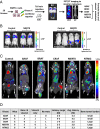
Screen of 20 kinases in human prostate cells identifies 5 kinases that drive bone and visceral metastasis. (A) Schema of the secondary screen. The 20 kinases identified in the primary screen were expressed in human prostate cells (RWPE-1 cells) and injected into the tail vein of mice. Immediately postinjection, mice were imaged by BLI to verify proper injection. Mice were monitored for metastasis by PET/CT imaging. (B) Representative BLI of mice injected with control or MERTK-expressing cells. At time (T) = 0, luciferase signal was detected in the lungs and, by T = 4 wk, luciferase signal was detected in the hind legs. (C) PET/CT images of mice injected with control cells or cells expressing the kinases ARAF, BRAF, CRAF, MERTK, and NTRK2. White arrows indicate anatomical sites of high glycolytic activity corresponding to sites of tumor growth. Scale bar on right corresponds to percent injected dose (ID) per gram (g) of tissue. (D) Table summarizing the outcomes of tail vein injections of RWPE-1 cells overexpressing ARAF, BRAF, CRAF, MERTK, and NTRK2. Listed are the number of mice tested per kinase, sites of metastatic colonization (“bone & visceral” or “visceral only”), latency (time point at which metastatic burden necessitated euthanasia), and tumor burden. The anatomical sites classified as visceral were lungs and lymph nodes. avg., average; M, month; mets, metastasis.
Fig. S3.
Fig. S3.
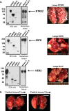
Identification of kinases promoting lung colonization when expressed in murine prostate cells. (A, Left) Western blot analysis of lung tissues showing the specific kinase that was found to be enriched in lung metastasis. (A, Right) Representative bright-field images of tumor nodules in lungs from the corresponding mice. (Scale bars, 5 mm.) (B) Bright-field images of lungs removed from mice 10 wk after injection with control Cap8 cells. (Scale bars, 5 mm.)
Fig. 4.
Fig. 4.
Histological analysis of bones recovered from mice injected with cells expressing ARAF, BRAF, CRAF, MERTK, and NTRK2 confirms that metastases are of human prostate epithelial cell origin. (Left two columns) H&E stains of the affected bones removed from mice injected with RWPE-1 cells expressing the five metastasis-promoting kinases. Images in Right five columns are 20× magnification of the area outlined by a black box in the first column. Tumor areas are outlined by black dotted lines and indicated by “T.” Bone and bone marrow are marked with “B” and “M,” respectively. (Right four columns) IHC staining of bone metastasis for overexpressed kinase, E-cadherin, HLA class I, and PSA. [Scale bars, 320 μm (Left) and 40 μm (Right five columns).]
Fig. S4.
Fig. S4.
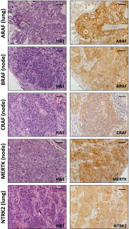
Overexpression of kinases in RWPE-1 cells drives the formation of bone and visceral metastases. Representative histology of visceral (lung or lymph node) metastases removed from mice injected with RWPE-1 cells expressing RAF family members, MERTK, or NTRK2. (Left) H&E images. (Right) Corresponding kinase-specific staining. (Scale bars, 40 μm.) The histology of bone metastases removed from the same mice is shown in Fig. 4.
Fig. S5.
Fig. S5.
Histological analysis of bones recovered from mice injected with cells expressing ARAF, BRAF, CRAF, MERTK, and NTRK2 confirms that metastases are of human prostate epithelial cell origin. (Left two columns) H&E stains of the affected bones removed from mice injected with RWPE-1 cells expressing the five metastasis-promoting kinases. Images in Right five columns are 20× magnification of the area outlined by a black box in the first column. (Right four columns) IHC staining of bone metastasis for overexpressed kinase, E-cadherin, HLA class I, and PSA. [Scale bars, 40 μm (Left) and 20 μm (Right five columns).]
Fig. 5.
Fig. 5.
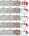
High levels of the five metastasis-promoting kinases are detected in human prostate cancer metastasis tissues. (Left) IHC staining for ARAF, BRAF, CRAF, MERTK, and NTRK2 in representative samples from TMAs containing tissue sections from normal prostate tissue, localized prostate cancer (Gleason 7–9), and metastatic prostate cancer. [Scale bars, 50 μm (large images) and 100 μm (small images).] (Right) Quantification of kinase expression in TMAs based on staining intensity. No immunoreactivity was scored as 0, whereas positive immunoreactivity was scored as 1 or 2 based on intensity. The distributions of scores between normal + metastatic tissues and localized + metastatic tissues were subjected to χ2 statistical analysis. Significance: *P ≤ 0.05, **P ≤ 0.01.

Comment in

References

    1. van Dodewaard-de Jong JM, et al. New treatment options for patients with metastatic prostate cancer: What is the optimal sequence? Clin Genitourin Cancer. 2015;13(4):271–279. - PubMed
    1. Nelson WG, De Marzo AM, Isaacs WB. Prostate cancer. N Engl J Med. 2003;349(4):366–381. - PubMed
    1. Bubendorf L, et al. Metastatic patterns of prostate cancer: An autopsy study of 1,589 patients. Hum Pathol. 2000;31(5):578–583. - PubMed
    1. de Bono JS, et al. TROPIC Investigators Prednisone plus cabazitaxel or mitoxantrone for metastatic castration-resistant prostate cancer progressing after docetaxel treatment: A randomised open-label trial. Lancet. 2010;376(9747):1147–1154. - PubMed
    1. Parker C, et al. ALSYMPCA Investigators Alpha emitter radium-223 and survival in metastatic prostate cancer. N Engl J Med. 2013;369(3):213–223. - PubMed

Publication types